Heart disease: Transgenic cell transplants offer hope against heart-attack after attacks
Transplants of genetically engineered cells could help to reduce the risk of fatal arrhythmia in the wake of a heart attack, according to new mouse studies. The discovery could offer a way to prevent ventricular tachycardia, a type of heart arrhythmia that is currently the main cause of sudden death in patients who have previously had a heart attack.
Previously, doctors have attempted to prevent ventricular tachycardia by implanting either bone marrow cells or other cells called skeletal myoblasts into heart tissue to help it recover from the damage sustained in a heart attack. But this approach has not proven successful because the cells never develop into true heart cells.
Bernd K. Fleischmann and colleagues report that, in mice, transplantation of cells called embryonic cardiomyocytes successfully reduces the danger of ventricular tachycardia. They also deduced that the key factor in these cells is a protein called connexin 43. When they genetically engineered skeletal myoblasts, which are more readily available, to express this same protein, they discovered that these cells were now equally effective in restoring heart function, thereby avoiding the need to use cells from embryos.
CONTACT
Bernd K. Fleischmann (University of Bonn, Germany)
Tel: +49 228 688 5200; E-mail: Bernd.Fleischmann@uni-bonn.de
Michael Kotlikoff (Cornell University, Ithaca, NY, USA) Co-author
Tel: +1 607 253 3771; E-mail: mik7@cornell.edu
Friday, December 07, 2007
Looking Through the Eyes of a Mouse, Scientists Monitor Circulating Cells in Its Bloodstream
Description
A team of researchers from the Wellman Center for Photomedicine at Massachusetts General Hospital (MGH) and Harvard Medical School (HMS) have developed an optical device that allows them to peer through the eyes of a mouse and monitor the cells passing through its bloodstream.
A team of researchers from the Wellman Center for Photomedicine at Massachusetts General Hospital (MGH) and Harvard Medical School (HMS) have developed an optical device that allows them to peer through the eyes of a mouse and monitor the cells passing through its bloodstream.
In the Dec. 1 issue of the journal Optics Letters, published by the Optical Society of America, the team describes how they used the device, called a retinal flow cytometer, to non-invasively sample the blood passing through the vessels in the retinal tissue in the back of the eye. There they were able to detect circulating fluorescently labeled cells as they wound their way through the mouse.
"We could detect and count circulating cells continuously without drawing blood samples," says MGH/HMS investigator Charles Lin.
The ability to count circulating cells is important in diseases like multiple myeloma because the number of cancer cells in the bloodstream at the onset of the disease may represent only a tiny fraction of circulating cells in the bloodstream.
Though few in number, these rare cells nevertheless can be relentless. Multiple myeloma starts when cancerous immune system cells residing in the bone marrow quickly multiply out of control. Rather than forming a solid tumor, though, they spread throughout the body and crowd out other cells in the bloodstream. Multiple myeloma cells can invade bones throughout the body, eroding and weakening them and leading to fractures and sometimes paralysis because of compression of the spinal cord. Eroding the bones can also drastically increase the calcium levels in the blood, sometimes causing kidney failure. Moreover, multiple myeloma cells can crowd out the oxygen-ferrying red blood cells in the bloodstream and cause anemia.
Multiple myeloma is treatable, but the disease has a high rate of recurrence. Scientists like Lin and his collaborator Irene Ghobrial of the Dana-Farber Cancer Institute are interested in helping people with multiple myeloma by finding better drugs and treatment strategies. The new optical device may become a valuable tool because it allows them to test the effect of various experimental chemotherapy agents and therapeutic strategies in mice with multiple myeloma.
Testing the effect of new chemotherapy agents in the bloodstream of a mouse has traditionally been difficult because there was no way to monitor the blood continuously. The best scientists could do was to draw blood samples at various time points and count the number of cancer cells in these samples. But looking for rare cancer cells required drawing a lot of blood. Repeated blood sampling from a single mouse was impossible, though, because mice only have a few milliliters of blood in their body.
A few years ago Lin and his colleagues devised a way to monitor cancer cells in rodents directly. They adapted a technique called flow cytometry, which scientists have used for decades to sort cells in the laboratory. Basically it involves streaming a complex mixture of cells in liquid past the focus of a powerful microscope and looking for particular cells—distinguishable because they have particular markers that are visible under the microscope.
Lin and his colleagues developed a way to use flow cytometry in vivo and monitor a mouse’s bloodstream directly. Initially they designed a device that could focus on a vessel in the ear of a mouse. While effective, this device was limited because the one tiny vessel in the mouse’s thin external ear did not have enough flow to adequately sample the bloodstream. It was a bit like trying to sample the traffic in New York by looking at a single side street in the Bronx. A better strategy would be to monitor several avenues at once—or in vascular terms, a cluster of several vessels at the same time.
This is exactly what the researchers are doing by looking into the back of a rodent’s eye. The retinal tissue is rich with blood supply, and Lin and his colleagues were able to sample a greater number of blood vessels and a much larger volume of blood. In initial feasibility experiments, they monitored a million circulating cells in the mouse that were fluorescently labeled with a marker that allowed them to be spotted with a microscope. The cytometer allowed them to track these cells as they trafficked though the bloodstream. They could observe approximately 250 cells per minute, and over time statistically model the flow of the cells.
This new device is not designed for humans and has not been tested in clinical trials. As a laboratory tool, however, it will allow the team to observe what happens when different chemotherapy agents are given to rodents—a standard early approach for evaluating the effectiveness of new chemotherapy agents and treatment strategies for humans.
The researchers are funded by grants from the National Institutes of Health.
Paper: "Retinal Flow Cytometer," by Clemens Alt et al., Optics Letters, Vol. 32, Issue 23, December 1, pp.3450-3452; abstract at http://www.opticsinfobase.org/abstract.cfm?URI=ol-32-23-3450.
Additional Reference: "Mechanisms of regulation of CXCR4/SDF-1 (CXCL12)–dependent migration and homing in multiple myeloma," by Yazan Alsayed et al., Blood, Vol. 109, Apr 2007, pp. 2708-2717.
About OSA
Uniting more than 70,000 professionals from 134 countries, the Optical Society of America (OSA) brings together the global optics community through its programs and initiatives. Since 1916 OSA has worked to advance the common interests of the field, providing educational resources to the scientists, engineers and business leaders who work in the field by promoting the science of light and the advanced technologies made possible by optics and photonics. OSA publications, events, technical groups and programs foster optics knowledge and scientific collaboration among all those with an interest in optics and photonics. For more information, visit http://www.osa.org.
Researchers Find Mechanism for Faulty Protein Disposal
Description
A discovery by St. Jude Children’s Research Hospital scientists offers new insights into how myeloma cells dispose of defective or excess proteins and could lead to new cancer treatments.
A discovery by St. Jude Children’s Research Hospital scientists offers new insights into how myeloma cells dispose of defective or excess proteins and could lead to new cancer treatments.
The researchers identified key cellular components that carry out protein disposal, a finding that helps to explain how cancer drugs called proteasome inhibitors interfere with this process. The discovery is important because the newly identified components of the protein disposal mechanism could be targets for novel cancer drugs designed to kill the cell by blocking this mechanism. A report on this work appears in the Nov. 30 issue of “Molecular Cell.”
Myelomas are cancers of plasma cells, which are the activated form of B lymphocytes—immune system cells that respond to infection by temporarily producing extremely large amounts of proteins called antibodies that attack the target. The rapidly multiplying cancer cells continually make large numbers of new antibodies, which increases the chance for errors in the production process, resulting in the accumulation of defective proteins that must be degraded.
“Proteasome inhibitors are currently being used to treat some types of cancer including multiple myelomas, although many aspects of this cellular process remain poorly understood,” said Linda Hendershot, Ph.D., a member of the St. Jude Department of Genetics and Tumor Cell Biology, and the paper’s senior author. “Our study sheds new light on how that process works.”
The St. Jude team focused their investigation on special channels called retrotranslocons in the membrane of the cell’s protein factory. The researchers also studied a small collection of molecules that pull defective proteins out of the factory through the retrotranslocon so they can be delivered to the cell’s protein shredder—a structure called the proteasome.
The protein factory, called the endoplasmic reticulum, is somewhat like an origami workshop: In the endoplasmic reticulum, molecules called chaperones help to fold up newly made proteins into the exact shape that enables that particular protein to perform its assigned task. Successfully folded proteins are transported to the cell surface or into the blood stream where they do their job. But if the folding does not occur or is faulty, defective proteins are ejected from the endoplasmic reticulum through channels called retrotranslocons and put into the proteasome, where they get degraded. This process, called endoplasmic reticulum-associated degradation, ensures that defective proteins do not accumulate in the endoplasmic reticulum and kill the cell by disrupting the vital process of protein folding.
Previous research identified a channel out of the endoplasmic reticulum for glycosylated proteins, or proteins tagged with sugar molecules. The channel relies on the detection of the sugar molecules to identify the proteins and send them to the proteasome. However, nothing was known about the disposal of non-glycosylated proteins (proteins with few or no sugar molecules attached). The St. Jude team found that this group of proteins exits the endoplasmic reticulum through a channel that is similar to the one used for glycoproteins but that has different components. The team focused the study on non-glycosylated proteins called light chains and heavy chains, which are the building blocks for antibodies made by plasma cells.
“We wanted to determine what happens to defective heavy and light chains in plasma cells so we could get a better understanding of the molecules and channels that allow these cells to get rid of defective proteins that can’t be used to make antibodies,” Hendershot said. When plasma cells become cancerous, they multiply rapidly and continue to produce large amounts of antibodies, some of which are not folded properly. These cells depend on endoplasmic reticulum-associated degradation to dispose of unwanted proteins before they clog up the endoplasmic reticulum and eventually kill the cells.
“The class of cancer drugs called proteasome inhibitors block endoplasmic reticulum-associated degradation as well as the destruction of proteins from other parts of the cells and cause defective proteins to overload this system,” she said. “We want to fully understand how endoplasmic reticulum-associated degradation works for antibodies made by plasma cells, so we can design more specific ways to block this process in myelomas.”
The St. Jude team first demonstrated that defective light chain and heavy chain proteins in plasma cells are degraded by the proteasome after being ejected from the endoplasmic reticulum and tagged with molecules called ubiquitin—a standard way the cell flags an unwanted protein for destruction.
The researchers then examined the menagerie of molecules that collaborate to pull defective proteins out of the endoplasmic reticulum and hand it over to the proteasome. Hendershot’s team showed previously that one of those molecules, a chaperone called BiP, initially helps newly made proteins undergo folding. If the folding operation fails, however, BiP becomes a conspirator with Herp, another member of the menagerie, to send the defective protein to the proteasome. Based on a series of detailed biochemical studies, the team showed that Herp binds to both the ubiquitinated protein and the proteasome, apparently serving as a bridge to direct the protein to the shredder.
In addition to BiP and Herp, three other members of the menagerie, Derlin-1, p97 and Hrd 1 collaborate with Herp to extract defective proteins from the retrotranslocon so Herp can hand it over to the proteasome.
“Our study shows for the first time the role Herp plays at the retrotranslocon,” said Yuki Okuda-Shimizu, Ph.D. a postdoctoral fellow in Hendershot’s laboratory who contributed significantly to the project. “The study also describes how non-glycosylated proteins are removed from the endoplasmic reticulum and disposed of. This information helps to explain how the process works and how we might design ways to block it in cancer cells.”
This work was supported by the National Institutes of Health, a Cancer Center Support Grant and ALSAC.(Newswise)
St. Jude Children's Research Hospital
St. Jude Children's Research Hospital is internationally recognized for its pioneering work in finding cures and saving children with cancer and other catastrophic diseases. Founded by late entertainer Danny Thomas and based in Memphis, Tenn., St. Jude freely shares its discoveries with scientific and medical communities around the world. No family ever pays for treatments not covered by insurance, and families without insurance are never asked to pay. St. Jude is financially supported by ALSAC, its fundraising organization.
How Ketamine ("Special K") Impairs Brain Circuitry
Description
Use of ketamine raising concerns by researchers at the UCSD School of Medicine who have found that ketamine leads to the impairments in brain circuitry observed in both drug abusers and schizophrenic patients by causing increased production of a toxic free radical called “superoxide.”
Scientists know that the drug ketamine – street name “Special K” – can induce schizophrenia-like symptoms in drug abusers. Ketamine is also used as an anesthetic and, more recently, as an antidepressant – raising concerns by researchers at the University of California, San Diego (UCSD) School of Medicine, who have found that ketamine leads to the impairments in brain circuitry observed in both drug abusers and schizophrenic patients by causing increased production of a toxic free radical called “superoxide.” Their findings, which could point the way to novel treatments for schizophrenia, will be published in the December 7 issue of the journal Science.
A research team led by Laura Dugan, M.D., Larry L. Hillblom Professor of Geriatrics and research scholar with the UCSD Stein Institute for Research on Aging, discovered an unexpected link between the inflammatory enzyme complex NADPH oxidase and the dysfunction of certain brain neurons exposed to ketamine. NADPH oxidase is normally found in white blood cells circulating outside the brain, where it helps kill bacterial and fungal infections by producing superoxide, a compound that can cause substantial damage to cells.
“Because of NADPH oxidase’s protective role in fighting infection, it was very surprising to find that the complex wears a second hat – it is also critical for modulating signaling in the brain,” said first author M. Margarita Behrens, Ph.D., Division of Geriatric Medicine, UCSD School of Medicine.
According to Behrens, it was known that ketamine initially impairs the inhibitory circuitry in the brain’s cortex and hippocampus by blocking the NMDA receptor, a molecule on the cell surface that controls the activity of neurons. But the UCSD researchers discovered that, as a result of blocking the receptor, ketamine also substantially increased the activity of NADPH oxidase, causing further disruption of neuronal signaling.
“Ketamine causes a ‘disinhibition’ of brain circuitry, taking the brakes off the system and causing overexcitation of the brain in response to a stimulus,” said Behrens. “This overexcitation activates NADPH oxidase, which then produces superoxide – resulting in detrimental changes in key synaptic proteins and profoundly affecting nervous system function.”
The result is impairment of the brain circuitry involved in memory, attention and other key functions related to learning. Loss of such functions sets up individuals for psychosis and deficits in information processing, resulting in symptoms such as hallucinations and delusions, as well as social withdrawal and cognitive problems, according to Behrens.
Using ketamine, Behrens and Dugan mimicked features of schizophrenia in mice, and then analyzed neurons in a region of the mouse brain that corresponds to the prefrontal cortex in humans where profound changes occur in patients with schizophrenia. The researchers found a substantial increase in the activity of NADPH oxidase, and that this activity made some neurons in this inhibitory circuitry “disappear.” When the researchers blocked the activity of NADPH oxidase with an inhibitor, or with a compound that annihilates superoxide, these neurons were protected.
“Our findings suggest that compounds that inhibit NADPH oxidase in the brain, without totally blocking its protective function of killing bacteria, could provide future therapies for schizophrenia or other diseases in humans that exhibit similar changes in neural circuitry,” said Behrens.
Additional contributors to the paper include Sameh S. Ali, Diep N. Dao, Jacinta Lucero, Grigoriy Shekhtman and Kevin L. Quick, Department of Medicine, UCSD Division of Geriatric Medicine. The research was funded in part by the Larry L. Hillblom Endowment and NARSAD.
One Mutation in Mosquito Virus Launched La Reunion Epidemic
Description
Researchers have discovered the key to how a mysterious mosquito-borne viral outbreak swept over the Indian Ocean island of La Reunion in 2005 and 2006, infecting about 266,000 people and causing at least 260 deaths — the first fatalities to be reported in connection with the virus, known as chikungunya.
Researchers have discovered the key to how a mysterious mosquito-borne viral outbreak swept over the Indian Ocean island of La Reunion in 2005 and 2006, infecting about 266,000 people and causing at least 260 deaths — the first fatalities to be reported in connection with the virus, known as chikungunya.
University of Texas Medical Branch at Galveston (UTMB) scientists proved that a single mutation in the virus glycoprotein E1 made it more infectious and facilitated its transmission by a mosquito species found on La Reunion and neighboring islands — one not previously involved in chikungunya outbreaks. The species, Aedes albopictus, is commonly known as the Asian tiger mosquito; it established itself in the United States about 20 years ago and has recently spread to southern Europe.
Chikungunya virus normally causes extreme arthritis-like joint pain, which can occasionally last for months or years. It takes its name from an African tribal word meaning “that which bends up,” a reference to the contorted, stooped posture some of its victims assume.
“Chikungunya virus had been known to be primarily carried by a different mosquito, Aedes aegypti, which is not found on La Reunion,” said UTMB professor Stephen Higgs, senior author of a paper on the discovery appearing online Dec. 7 in PloS Pathogens, a peer-reviewed open-access journal published by the Public Library of Science. “Adaptation to Aedes albopictus and introduction to a human population that had never been exposed to the virus before set everything up for this outbreak.”
Numerous tourists became infected, many of whom carried chikungunya home with them. Although the La Reunion strain of the virus did not cause a subsequent outbreak in Europe, Higgs said, it could have done so. In fact, another strain of the virus responsible for an ongoing outbreak in India has spread to Aedes albopictus mosquitoes and humans in Italy.
The UTMB researchers focused their attention on a single-amino acid change in the La Reunion chikungunya strain and found that this E1 A226V mutation led to increased virus infectivity for Aedes albopictus. To further prove the role of this mutation, lead author and graduate student Konstantin Tsetsarkin inserted that change into a chikungunya strain collected in West Africa in 1983, and then showed that it dramatically increased the virus’ ability to infect Aedes albopictus.
“After we finished with the infectivity experiments, we analyzed the ability of the virus to disseminate to mosquito salivary glands and then be transmitted by mosquitoes,” Tsetsarkin said. “Then we simultaneously infected Aedes albopictus mosquitoes with equal amounts of the mutant virus and the original strain, and we saw that the virus with the E1 A226V mutation was predominately transmitted over the other one — the mutation gave the La Reunion strain an evolutionary advantage in Aedes albopictus, enabling it to out-compete the other strain.”
The key mutation, Tsetsarkin said, was linked to the virus’ dependence on cholesterol in mosquito cell membranes, a key factor in the virus’ ability to infect those cells and spread to mosquito salivary glands. Like other mosquito-borne viruses, chikungunya spreads to humans in mosquito saliva when the insects bite people.
“This is such a simple genetic change — the equivalent of a missing comma in a six-page short story — and yet it facilitated a huge epidemic,” Higgs said. “With Aedes albopictus already present in so many countries and likely to spread to others, perhaps helped by global warming, this shows us that we need to be ready for the possibility that chikungunya will soon be spreading to other locations as well.”
Tuesday, November 27, 2007
High-Performance Transistors Produced from Carbon 60
Description
Using room-temperature processing, Georgia Tech researchers have fabricated high-performance field effect transistors with thin films of Carbon 60, also known as fullerene. The work represents another milestone toward practical applications for large area, low-cost electronic circuits on flexible organic substrates.
Using room-temperature processing, researchers at the Georgia Institute of Technology have fabricated high-performance field effect transistors with thin films of Carbon 60, also known as fullerene. The ability to produce devices with such performance with an organic semiconductor represents another milestone toward practical applications for large area, low-cost electronic circuits on flexible organic substrates.
The new devices – which have electron-mobility values higher than amorphous silicon, low threshold voltages, large on-off ratios and high operational stability – could encourage more designers to begin working on such circuitry for displays, active electronic billboards, RFID tags and other applications that use flexible substrates.
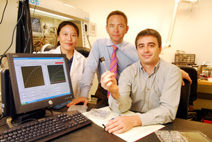
“If you open a textbook and look at what a thin-film transistor should do, we are pretty close now,” said Bernard Kippelen, a professor in Georgia Tech’s School of Electrical and Computer Engineering and the Center for Organic Photonics and Electronics. “Now that we have shown very nice single transistors, we want to demonstrate functional devices that are combinations of multiple components. We have everything ready to do that.”
Fabrication of the C60 transistors was reported in the journal Applied Physics Letters on August 27th. The research was supported by the U.S. National Science Foundation through the STC program MDITR, and the U.S. Office of Naval Research.
Researchers have been interested in making field-effect transistors and other devices from organic semiconductors that can be processed onto various substrates, including flexible plastic materials. As an organic semiconductor material, C60 is attractive because it can provide high electron mobility – a measure of how fast current can flow. Previous reports have shown that C60 can yield mobility values as high as six square centimeters per volt-second (6 cm2/V/s). However, that record was achieved using a hot-wall epitaxy process requiring processing temperatures of 250 degrees Celsius – too hot for most flexible plastic substrates.
Though the transistors produced by Kippelen’s research team display slightly lower electron mobility – 2.7 to 5 cm2/V/s – they can be produced at room temperature.
“If you want to deposit transistors on a plastic substrate, you really can’t have any process at a temperature of more than 150 degrees Celsius,” Kippelen said. “With room temperature deposition, you can be compatible with many different substrates. For low-cost, large area electronics, that is an essential component.”
Because they are sensitive to contact with oxygen, the C60 transistors must operate under a nitrogen atmosphere. Kippelen expects to address that limitation by using other fullerene molecules – and properly packaging the devices.
The new transistors were fabricated on silicon for convenience. While Kippelen isn’t underestimating the potential difficulty of moving to an organic substrate, he says that challenge can be overcome.
Though their performance is impressive, the C60 transistors won’t threaten conventional CMOS chips based on silicon. That’s because the applications Kippelen has in mind don’t require high performance.
“There are a lot of applications where you don’t necessarily need millions of fast transistors,” he said. “The performance we need is by far much lower than what you can get in a CMOS chip. But whereas CMOS is extremely powerful and can be relatively low in cost because you can make a lot of circuits on a wafer, for large area applications CMOS is not economical.”
A different set of goals drives electronic components for use with low-cost organic displays, active billboards and similar applications.
“If you look at a video display, which has a refresh rate of 60 Hz, than means you have to refresh the screen every 16 milliseconds,” he noted. “That is a fairly low speed compared to a Pentium processor in your computer. There is no point in trying to use organic materials for high-speed processing because silicon is already very advanced and has much higher carrier mobility.”
Now that they have demonstrated attractive field-effect C60 transistors, Kippelen and collaborators Xiao-Hong Zhang and Benoit Domercq plan to produce other electronic components such as inverters, ring oscillators, logic gates, and drivers for active matrix displays and imaging devices. Assembling these more complex systems will showcase the advantages of the C60 devices.
“The goal is to increase the complexity of the circuits to see how that high mobility can be used to make more complex structures with unprecedented performance,” Kippelen said.
The researchers fabricated the transistors by depositing C60 molecules from the vapor phase into a thin film atop a silicon substrate onto which a gate electrode and gate dielectric had already been fabricated. The source and drain electrodes were then deposited on top of the C60 films through a shadow mask.
Kippelen’s team has been working with C60 for nearly ten years, and is also using the material in photovoltaic cells. Beyond the technical advance, Kippelen believes this new work demonstrates the growing maturity of organic electronics.
“This progress may trigger interest among more conventional electronic engineers,” he said. “Most engineers would like to work with the latest technology platform, but they would like to see a level of performance showing they could actually implement these circuits. If you can demonstrate – as we have – that you can get transistors with good reproducibility, good stability, near-zero threshold voltages, large on-off current ratios and performance levels higher than amorphous silicon, that may convince designers to consider this technology.”
(Newswise)

Description
Using room-temperature processing, Georgia Tech researchers have fabricated high-performance field effect transistors with thin films of Carbon 60, also known as fullerene. The work represents another milestone toward practical applications for large area, low-cost electronic circuits on flexible organic substrates.
Using room-temperature processing, researchers at the Georgia Institute of Technology have fabricated high-performance field effect transistors with thin films of Carbon 60, also known as fullerene. The ability to produce devices with such performance with an organic semiconductor represents another milestone toward practical applications for large area, low-cost electronic circuits on flexible organic substrates.
The new devices – which have electron-mobility values higher than amorphous silicon, low threshold voltages, large on-off ratios and high operational stability – could encourage more designers to begin working on such circuitry for displays, active electronic billboards, RFID tags and other applications that use flexible substrates.
“If you open a textbook and look at what a thin-film transistor should do, we are pretty close now,” said Bernard Kippelen, a professor in Georgia Tech’s School of Electrical and Computer Engineering and the Center for Organic Photonics and Electronics. “Now that we have shown very nice single transistors, we want to demonstrate functional devices that are combinations of multiple components. We have everything ready to do that.”
Fabrication of the C60 transistors was reported in the journal Applied Physics Letters on August 27th. The research was supported by the U.S. National Science Foundation through the STC program MDITR, and the U.S. Office of Naval Research.
Researchers have been interested in making field-effect transistors and other devices from organic semiconductors that can be processed onto various substrates, including flexible plastic materials. As an organic semiconductor material, C60 is attractive because it can provide high electron mobility – a measure of how fast current can flow. Previous reports have shown that C60 can yield mobility values as high as six square centimeters per volt-second (6 cm2/V/s). However, that record was achieved using a hot-wall epitaxy process requiring processing temperatures of 250 degrees Celsius – too hot for most flexible plastic substrates.
Though the transistors produced by Kippelen’s research team display slightly lower electron mobility – 2.7 to 5 cm2/V/s – they can be produced at room temperature.
“If you want to deposit transistors on a plastic substrate, you really can’t have any process at a temperature of more than 150 degrees Celsius,” Kippelen said. “With room temperature deposition, you can be compatible with many different substrates. For low-cost, large area electronics, that is an essential component.”
Because they are sensitive to contact with oxygen, the C60 transistors must operate under a nitrogen atmosphere. Kippelen expects to address that limitation by using other fullerene molecules – and properly packaging the devices.
The new transistors were fabricated on silicon for convenience. While Kippelen isn’t underestimating the potential difficulty of moving to an organic substrate, he says that challenge can be overcome.
Though their performance is impressive, the C60 transistors won’t threaten conventional CMOS chips based on silicon. That’s because the applications Kippelen has in mind don’t require high performance.
“There are a lot of applications where you don’t necessarily need millions of fast transistors,” he said. “The performance we need is by far much lower than what you can get in a CMOS chip. But whereas CMOS is extremely powerful and can be relatively low in cost because you can make a lot of circuits on a wafer, for large area applications CMOS is not economical.”
A different set of goals drives electronic components for use with low-cost organic displays, active billboards and similar applications.
“If you look at a video display, which has a refresh rate of 60 Hz, than means you have to refresh the screen every 16 milliseconds,” he noted. “That is a fairly low speed compared to a Pentium processor in your computer. There is no point in trying to use organic materials for high-speed processing because silicon is already very advanced and has much higher carrier mobility.”
Now that they have demonstrated attractive field-effect C60 transistors, Kippelen and collaborators Xiao-Hong Zhang and Benoit Domercq plan to produce other electronic components such as inverters, ring oscillators, logic gates, and drivers for active matrix displays and imaging devices. Assembling these more complex systems will showcase the advantages of the C60 devices.
“The goal is to increase the complexity of the circuits to see how that high mobility can be used to make more complex structures with unprecedented performance,” Kippelen said.
The researchers fabricated the transistors by depositing C60 molecules from the vapor phase into a thin film atop a silicon substrate onto which a gate electrode and gate dielectric had already been fabricated. The source and drain electrodes were then deposited on top of the C60 films through a shadow mask.
Kippelen’s team has been working with C60 for nearly ten years, and is also using the material in photovoltaic cells. Beyond the technical advance, Kippelen believes this new work demonstrates the growing maturity of organic electronics.
“This progress may trigger interest among more conventional electronic engineers,” he said. “Most engineers would like to work with the latest technology platform, but they would like to see a level of performance showing they could actually implement these circuits. If you can demonstrate – as we have – that you can get transistors with good reproducibility, good stability, near-zero threshold voltages, large on-off current ratios and performance levels higher than amorphous silicon, that may convince designers to consider this technology.”
(Newswise)
Monday, November 19, 2007
NEUROSCIENCE : Optimistic neurons
In rats given a choice between two rewards, neurons that predict the value of expected rewards responded as though the animal had chosen the best available reward, no matter what the rat actually did. These results suggest that the opportunity to make a choice may be as valuable as the best available option on the menu.
The authors trained rats to learn to associate each of three odours with an action. Two of the odours signalled that the rat was required to move either left or right to receive a reward, with one location containing a better reward than the other. The third odour indicated that the animal could choose to go to either location. Dopaminergic neurons in the ventral tegmental area, part of the brain’s “reward processing system,” responded more for the odour that cued the rat to move to the location associated with the better reward, as expected from previous studies.
The rewards varied either in the amount of juice or in how long the rat had to wait for its delivery, and responses to the odour cues were correlated with both reward size and delay. The dopaminergic neurons responded just as strongly to the odour indicating that the rat could choose which reward to collect as to the better reward, even though the rat eventually chose the worse reward nearly 30% of the time on the free choice trials.
Author Contact:
Matthew Roesch (University of Maryland School of Medicine, Baltimore, MD, USA)
Tel: +1 410 706 8910; E-mail: mroes001@umaryland.edu
GENETICS : Gene prevents sudden death in mice after infection
Mice that lack a particular gene die suddenly and without overt signs of illness in response to an infection that is usually harmless, according to a study. This work may lead to new insights into the origins of sudden death in humans, although such a link has not yet been made.
Bruce Beutler and colleagues treated mice with a chemical mutagen – an agent that changes genetic information; and examined the third-generation offspring of the mutant mice for susceptibility to cytomegalovirus (CMV). This virus, at the dose delivered, is normally harmless. The progeny of four of the original mutants, however, died suddenly between 36 hours and 3 days after inoculation. One of these lines has a large deletion in Kcnj8, a gene encoding a component of a potassium channel expressed in smooth muscle and endothelial cells of the coronary artery (two of the other lines carry different mutations in Kcnj8).
The protein that interacts with Kcnj8 has a counterpart in the fruit fly, and the authors show that it similarly protects flies against sudden death after challenge with flock house virus (FHV). The authors propose that this potassium channel is required for the coronary arteries to survive the systemic metabolic stress and arterial constriction that accompanies the innate immune response to viruses such as CMV and FHV.
Author contact:
Bruce Beutler (Scripps Research Institute, La Jolla, CA, USA)
Tel: +1 858 784 8610; E-mail: bruce@scripps.edu
PHYSICS : Neural networks organise themselves
Neural networks dynamically organize themselves to operate in a range that is optimal for information processing, according to a theoretical model. The results provide an understanding of how neurons interact with each other and how they can build efficient networks.
Neurons signal to each other through junctions known as synapses. Using these connections they can build extended networks. Computer simulations of neural networks indicate that for specific connection patterns, properties such as computational power or memory capacity are maximized. This picture is supported by experimental findings in cell cultures. However, how neural networks can be tuned to the optimal setting is still an open question.
Michael Herrmann and colleagues argue that there is no need for fine-tuning. They factor in that synapses are not static — that is, the efficiency of transmission through synapses depends on the frequency of their use — and show in their model that neural networks dynamically organize themselves to operate in a favourable range.
Author contact:
Michael Herrmann (Universität Göttingen, Germany)
Tel: +49 551 517 6424; E-mail: michael@nld.ds.mpg.de
Immunity: Holding dormant cancers in check
Dormant cancer cells are actively kept in check by the host’s immune system — those that escape go on to develop into clinically detectable tumours. A paper identifies a crucial stage in the battle, at which point defences stall the expansion of cancer cells that may have managed to dodge past early immunosurveillance.
Robert Schreiber and co-workers use a mouse model to show that the animal’s immune system can keep tumour growth in check over an extended period. Clinicians have suspected the existence of such an ‘equilibrium’ state, because dormant cancers sometimes take off when inadvertently transferred from a donor to an immunosuppressed recipient during organ transplantation.
This newly discovered staging post could also explain the presence of occult tumour cells — in the prostate, for example — in individuals with no symptoms of disease. Eventually it could be used to devise immunotherapies for tightening control of tumour growth, suggest the authors.
In an accompanying News & Views article, Cornelis Melief comments on the ‘startling results’ and says: ‘they demonstrate that considering cancer as a fatal disease is not always appropriate’.
Author contact:
Robert Schreiber (Washington University School of Medicine, St Louis, MO, USA)
Tel: +1 314 362 8747; E-mail: schreiber@immunology.wustl.edu
Cornelis Melief (Leiden University Medical Center, Netherlands) N&V author
Tel: +31 71 526 3800; E-mail: C.Melief@lumc.nl
Monday, November 12, 2007
STRUCTURAL AND MOLECULAR BIOLOGY : Stalling chemotherapy damage
The understanding of how healthy cells cope with damages caused by anti-cancer drugs is furthered by new findings.
Transcription is the process whereby genetic information is transferred from DNA to RNA, in most cases leading to production of a particular protein. DNA damage, such as that caused by some anticancer drugs, can lead to errors in the RNA produced during transcription, resulting in incorrect protein production that may be harmful to the cell.
Patrick Cramer and colleagues have investigated how transcription machinery avoids DNA lesions caused by cisplatin, a widely used chemotherapy drug. They found that the cisplatin lesion forces the transcription machinery to stop before it reaches the lesion. This transcriptional “stalling” triggers a DNA-repair pathway that can remove the toxic lesion.
Author contact:
Patrick Cramer (Gene Center Munich, Germany)
Tel: +49 89 2180 76965; e-mail: cramer@LMB.uni-muenchen.de
METHODS : Controlling protein stability in parasites
Methods to regulate protein expression in two hazardous parasites. This research will provide valuable tools for understanding disease development.
Toxoplasma gondii, a parasite that can cause encephalitis and neurological diseases, and Plasmodium falciparum, a malaria parasite, have both had their genome sequenced. Still lacking are methods to control protein expression on a large scale, so the effects of proteins on parasite biology and pathogenesis can be studied.
The research groups of Daniel Goldberg and Markus Meissner adapted a system, originally developed in mammalian cells, that allows them to trigger the degradation of any protein at will. The only pre-requisite is that the protein is coupled to a short peptide that makes protein stability dependent on the presence of another component, appropriately named Shield. If Shield is added to the parasites the targeted protein is stable, but if Shield is withdrawn, the protein is degraded and the effect of its loss on the parasite can be studied.
This fast and efficient method for regulating protein levels will allow a genome wide analysis of their roles in the parasite life cycle and the interaction with its host.
Author contacts:
Markus Meissner (University Hospital Heidelberg, Heidelberg, Germany)
Tel: +49 6221 566518; E-mail: markus.meissner@med.uni-heidelberg.de
Daniel Goldberg (Washington University School of Medicine, St. Louis, MO, USA)
Tel: +1 314 362 1514; E-mail: goldberg@borcim.wustl.edu
Getting to the root of a developmental mystery
Researchers have revealed how two closely related proteins trigger opposing effects in developing roots
The formation of root epidermis in Arabidopsis thaliana, a popular plant research model, offers a valuable means for studying cell differentiation in developing tissues. During root development, progenitor cells yield two classes of epidermal cells, hair cells and hairless cells, which form in a fixed pattern along the root.
Previous research has identified factors that determine whether hair cells or hairless cells form. Two of the genes involved, CAPRICE (CPC) and WEREWOLF (WER), encode closely related transcription factors that exhibit notable functional differences, which piqued the interest of Takuji Wada, a researcher at the RIKEN Plant Sciences Center in Yokohama. “CPC activates root-hair cell differentiation whereas WER represses it, even though both belong to the same family of transcription factors,” explains Wada, “so I wondered why these two factors have opposite effects.”
CPC and WER belong to the MYB family of transcription factors, whose distinguishing characteristics include several domains with repeated amino acid sequences. Wada and his colleagues generated several CPC and WER variants, swapping different portions of one of these repeat domains (Myb R3) between the two proteins. These were expressed in plant strains that lack functional CPC or WER in order to understand the relevant regions that determine each protein’s function1.
Wada’s team found that WER only inhibited hair cell formation when its entire R3 domain was intact. On the other hand, most of CPC’s R3 domain could be replaced without impeding its activity (Fig. 1). Subsequent experiments showed that both proteins bind common targets—GL3 and EGL3, two proteins that induce hairless cell formation. Myb R3 substitutions had no effect on this activity, but did affect the ability of WER to bind DNA—a property absent in CPC. “The sequence of the WER MYB R3 domain is restricted—the equivalent domain of CPC cannot be substituted for it,” says Wada. “Therefore, these restricted sequences are necessary for binding to DNA.”
Wada’s group believes that both WER and CPC compete for binding GL3 and EGL3. When WER binds, its unique DNA-binding sequences allow it to recruit these proteins in order to regulate genes responsible for hairless cell formation. However, when CPC is present as a competitor, no DNA binding takes place and hair cells develop instead. Based on the findings from this study, Wada suggests that CPC probably originated from a duplicate copy of the WER gene, a truncated younger sibling that nevertheless evolved into an effective rival.
Reference
1. Tominaga, R., Iwata, M., Okada, K. & Wada, T. Functional analysis of the epidermal-specific MYB genes CAPRICE and WEREWOLF in Arabidopsis. Plant Cell 19, 2264–2277 (2007).
Cell fusion may create niche for immune cell education
Japanese researchers have identified a subset of cells they believe may induce the formation of a network of follicular dendritic cells (FDC) in the spleen and lymph nodes.
Researchers identify possible precursors of lymphoid tissue cellular network
Japanese researchers have identified a subset of cells they believe may induce the formation of a network of follicular dendritic cells (FDC) in the spleen and lymph nodes.
A recent paper by Hiroshi Ohno and colleagues at the RIKEN Research Center for Allergy and Immunology, Yokohama, suggests that in the mouse, spleen cells expressing the cell surface marker proteins CD35, involved in processing and clearance of immune complexes, and B220, found on almost all immune system cells, can induce the formation of these networks and ultimately lymphoid follicles1 (Fig. 1).
These so-called FDC form a reticular network of cells in the spleen and lymph nodes that trap immune complexes of antibodies, antigens and associated molecules. The network plays a critical role in the development and maturation of the antibody-producing B lymphocytes (B cells). If a B cell binds weakly to an antigen trapped on the surface of an FDC, it undergoes programmed cell death (apoptosis). On the other hand, a B cell that has a high affinity for the trapped antigen survives to become an antibody-producing plasmablast, and ultimately a memory B cell. This is the fundamental process that underpins the ability of the immune system to respond quickly to attack by pathogenic infection.
But it is not simply a matter of recognition; it appears that a complex interaction of cells and molecules and cellular architecture within the dynamic microenvironment of the lymphoid follicle is required for B cell maturation. The players include connective tissue cells called stromal cells.
“The intrigue of the lymphoid follicle stems from the complexity of its microarchitecture, comprising immune cells and stromal cells, adhesion molecules, cytokines and antigen-antibody complexes, and the relationships between these components, in the formation of B cell-follicular dendritic cell aggregates and the regulation of B cell differentiation,” says Takaya Murakami, the first author of the paper.
Results from a series of experiments both in vitro and in vivo suggest that the splenic cells with the CD35 and B220 proteins on their surface (CD35+B220+ cells) interact with stromal cells to create a niche for migrating B cells, forming cell clusters. The researchers believe that this may play a critical role in FDC network development and the subsequent formation of lymphoid follicles. There is also some evidence that the stromal cells may fuse with the CD35+B220+ cells during this process.
Further investigation of the role of stromal cells in the development of the lymphoid follicles and B cell maturation is planned, says Murakami.
Reference
1. Murakami, T., Chen, X., Hase, K., Sakamoto, A., Nishigaki, C. & Ohno, H. Splenic CD19-CD35+B220+ cells function as an inducer of follicular dendritic cell network formation. Blood 110, 1215–1224 (2007).
Japanese researchers have identified a subset of cells they believe may induce the formation of a network of follicular dendritic cells (FDC) in the spleen and lymph nodes.
Researchers identify possible precursors of lymphoid tissue cellular network
Japanese researchers have identified a subset of cells they believe may induce the formation of a network of follicular dendritic cells (FDC) in the spleen and lymph nodes.
A recent paper by Hiroshi Ohno and colleagues at the RIKEN Research Center for Allergy and Immunology, Yokohama, suggests that in the mouse, spleen cells expressing the cell surface marker proteins CD35, involved in processing and clearance of immune complexes, and B220, found on almost all immune system cells, can induce the formation of these networks and ultimately lymphoid follicles1 (Fig. 1).
These so-called FDC form a reticular network of cells in the spleen and lymph nodes that trap immune complexes of antibodies, antigens and associated molecules. The network plays a critical role in the development and maturation of the antibody-producing B lymphocytes (B cells). If a B cell binds weakly to an antigen trapped on the surface of an FDC, it undergoes programmed cell death (apoptosis). On the other hand, a B cell that has a high affinity for the trapped antigen survives to become an antibody-producing plasmablast, and ultimately a memory B cell. This is the fundamental process that underpins the ability of the immune system to respond quickly to attack by pathogenic infection.
But it is not simply a matter of recognition; it appears that a complex interaction of cells and molecules and cellular architecture within the dynamic microenvironment of the lymphoid follicle is required for B cell maturation. The players include connective tissue cells called stromal cells.
“The intrigue of the lymphoid follicle stems from the complexity of its microarchitecture, comprising immune cells and stromal cells, adhesion molecules, cytokines and antigen-antibody complexes, and the relationships between these components, in the formation of B cell-follicular dendritic cell aggregates and the regulation of B cell differentiation,” says Takaya Murakami, the first author of the paper.
Results from a series of experiments both in vitro and in vivo suggest that the splenic cells with the CD35 and B220 proteins on their surface (CD35+B220+ cells) interact with stromal cells to create a niche for migrating B cells, forming cell clusters. The researchers believe that this may play a critical role in FDC network development and the subsequent formation of lymphoid follicles. There is also some evidence that the stromal cells may fuse with the CD35+B220+ cells during this process.
Further investigation of the role of stromal cells in the development of the lymphoid follicles and B cell maturation is planned, says Murakami.
Reference
1. Murakami, T., Chen, X., Hase, K., Sakamoto, A., Nishigaki, C. & Ohno, H. Splenic CD19-CD35+B220+ cells function as an inducer of follicular dendritic cell network formation. Blood 110, 1215–1224 (2007).
Wednesday, November 07, 2007
Dependence on Illicit and Licit DrugsGenes Play Role in Risk for
Description
The genes that play a role in illegal drug abuse are not entirely the same as those involved in dependence on legal substances like alcohol and nicotine, and caffeine addiction appears to be genetically independent of all the others, according to a study led by Virginia Commonwealth University researchers.
Research Highlights:
• Genes that play a role in illegal drug abuse are not entirely the same as those involved in dependence on legal substances like alcohol and nicotine
• Caffeine addiction appears to be genetically independent of the others
• Findings could guide efforts to localize genes that influence risk for psychoactive drug abuse or dependence
• First study to examine across the sexes and the degree to which risk factors for dependence are shared between illicit and licit drugs
The genes that play a role in illegal drug abuse are not entirely the same as those involved in dependence on legal substances like alcohol and nicotine, and caffeine addiction appears to be genetically independent of all the others, according to a study led by Virginia Commonwealth University researchers.
The findings may guide efforts by researchers to use molecular genetic tools to localize genes that influence risk for psychoactive drug abuse or dependence, or A/D.
In the November issue of the Archives of General Psychiatry, a journal of the American Medical Association, researchers examined the degree to which genetic and environmental risk factors for dependence were shared between illicit and the more commonly used licit psychoactive drugs among men and women.
“We wanted to know whether there was a single set of genes that influence risk for A/D on all substances,” said Kenneth S. Kendler, M.D., a professor of psychiatry and human genetics in VCU’s School of Medicine and lead author on the study.
“Our findings suggested two genetic factors - one which strongly impacted on risk for A/D of illicit drugs, such as cannabis and cocaine, and one that impacted on risk for A/D of licit drugs, including caffeine, nicotine and alcohol. However, these two factors were rather strongly correlated,” he said. “It was also of interest to note that the genes for caffeine A/D were pretty independent of those found for all the other substances.”
Kendler and his colleagues examined lifetime symptoms of abuse of and dependence on marijuana, cocaine, alcohol, caffeine and nicotine among 4,865 male-male and female-female twin pairs through a series of personal interviews. The data collected from the interviews was analyzed using the methods of structural equation modeling.
The twin pairs that participated were from the Virginia Adult Twin Study of Psychiatric and Substance Use Disorders. These twin pairs were ascertained from the Virginia Twin Registry. The Virginia Twin Registry, now part of the VCU Mid-Atlantic Twin Registry, contains a population-based record of twins from Virginia, North Carolina and South Carolina.
“This study also confirmed the strong role that genetic factors play in influencing our vulnerability to drug abuse and dependence,” Kendler said. Heritability – the proportion of individual differences in risk due to genetic differences – was estimated in this study to be more than 70 percent for cocaine, cannabis and nicotine A/D, nearly 60 percent for alcohol A/D, and, interestingly, quite a bit lower – around 35 percent – for caffeine A/D, he said.
In previous studies, researchers examined an array of illegal substances and did not include commonly used licit drugs, and included only male participates. This was the first study of its kind to examine across the sexes and degree to which risk factors for dependence were shared between illicit and licit drugs.
This work was supported by grants from the National Institutes of Health.
Kendler collaborated with John Meyer, M.S., from the Department of Psychiatry at VCU; and Carol A. Prescott, Ph.D., from the Department of Psychology, University of Southern California.
About VCU and the VCU Medical Center: Virginia Commonwealth University is the largest university in Virginia and ranks among the top 100 universities in the country in sponsored research. Located on two downtown campuses in Richmond, VCU enrolls more than 30,000 students in nearly 200 certificate and degree programs in the arts, sciences and humanities. Sixty-three of the programs are unique in Virginia, many of them crossing the disciplines of VCU’s 15 schools and one college. MCV Hospitals and the health sciences schools of Virginia Commonwealth University compose the VCU Medical Center, one of the nation’s leading academic medical centers. For more, see http://www.vcu.edu. (Newswise)
Thursday, November 01, 2007
Caribbean reefs struggling to recover from ecological sucker punches
A new study highlights the series of setbacks that have beset Caribbean coral reefs over the past few decades. The study charts the combination of events that has left the coral reefs in danger of converting to a new ecosystem type — one dominated not by living coral but by blooms of algae.
A group of ecologists led by Peter J. Mumby constructed a mathematical model to investigate the effects of a series of events, each of which acted as sucker punches that damaged reef health over the 1980s. First, coral was damaged by Hurricane Allen in 1980, and shortly after, in 1983, the ecosystem was rocked by a mass die-off of the urchin Diadema antillarum, which help to maintain coral health by grazing algae growing on dead coral, allowing the reef to be recolonized by healthy new coral colonies.
As Mumby and colleagues explain this combination of events — as well as the impact of the immense Hurricane Gilbert in 1988 — left the coral vulnerable to being taken over by algae. Their analysis may offer strategies for rescuing the reef ecosystem, perhaps by promoting populations of parrotfish, which also graze algae growing on the reef.
CONTACT
Peter J. Mumby (University of Exeter, UK)
Climate: Fires affect carbon balance in boreal forest
Carbon balance in the Canadian boreal forest is driven primarily by fire disturbance rather than by climate change. The researchers claim that overall changes in climate have not yet been felt in this large boreal region, and that variations in the landscape carbon balance and vegetation dominance have so far been largely driven by increases in fire frequency.
Ben Bond-Lamberty and colleagues use a process model with three competing vegetation types to examine the effects of climate, carbon dioxide concentrations and fire disturbance on a large area of Canadian boreal forest. They report that the carbon balance between 1948 and 2005 was driven largely by changes in fire disturbance. More frequent and larger fires in the late twentieth century resulted in deciduous trees and mosses increasing production at the expense of coniferous trees. They also find that poor soil drainage decreased the variability of the landscape carbon balance, suggesting that increased climate and hydrological changes do have the potential to affect disproportionately the carbon dynamics of these areas.
CONTACT
Ben Bond-Lamberty (University of Wisconsin-Madison, Madison, WI, USA)
Tel: +1 608 265-5628; E-mail: bpbond@wisc.edu
Neuroscience: The brain in glorious Technicolor
With a combination of genetic tricks and fancy proteins, researchers have colourfully labelled hundreds of individual neurons with distinctive hues to create a ‘Brainbow’. The research, takes mapping to a new level, and results in the labelling of neurons with approximately 90 different colour combinations.
Over a hundred years ago, Ramon Y Cajal’s use of Golgi staining on nerve cells opened the gates to modern neuroscience, but until now it has been tough to map out individual cells in each neuronal circuit. Jeff Lichtman and colleagues have developed the Technicolor version of Golgi staining, Brainbow, allowing more detailed reconstructions of brain circuits. This provides a key step towards modelling how the nervous system works normally and in diseased brains.
CONTACT
Jeff Lichtman (Harvard University, Cambridge, Ma, USA)
Tel: +1 617 496 8943; E-mail: jeff@mcb.harvard.edu
Alternative rewards may reduce addiction
Nondrug rewards could be a viable alternative to addictive drugs according to a study published in Neuropsychopharmacology. This finding could have important implications for those seeking treatment for drug addiction.
Serge Ahmed and colleagues found that rats with the hallmarks of heroin addiction used the substance less when a nondrug reward in the form of food was available. They also found that more dependant rats responded more to this alternative reward. This research indicates that the addicted brain remains reactive to nondrug rewards, and challenges the belief that the transition to addiction is associated with an individual valuing a drug more and nondrug rewards less. While studies in humans are needed, this finding suggests that heroin addiction could potentially be alleviated by increasing the availability of nondrug rewards to those with an addiction.
Contact:
Serge Ahmed (Université Victor-Segalen Bordeaux 2, Bordeaux, France)
Tel: +33 557 571 566; E-mail: sahmed@u-bordeaux2.fr
Evolution: Outside-in?
Theories that explain how the ‘primordial soup’ from which the first cell formed are challenged by a new model presented by Gareth Griffiths.
The theory that all life evolved from one source — the last common ancestor — as proposed by Charles Darwin and others, enjoys considerable support among evolutionary biologists. But questions remain about how the features that comprise a cell — membrane, cytoskeleton, genetic material — evolve to form a functional cell.
Numerous models propose that the formation of cells occurred inside randomly formed compartments (called vesicles) by a process known as the ‘cytoplasm within the vesicle’ scenario. But experimental validation of this theory is limited. Gareth Griffiths now describes an alternative theory that challenges previous dogma: the ‘cytoplasm outside the vesicle’ scenario. This theory states that early cytoplasmic components evolved in fresh water, probably on a mineral rock surface that also anchored small vesicles upon which the cytoplasm evolved.
The crucial difference is that this model proposes that the cytoskeleton, which maintains the three-dimensional structure of a cell and is involved in diverse cellular functions such as motility and transport, is required before, and as part of, the mechanism that led to cell formation. This theory provides an explanation to some of the mystifying questions in evolution, such as how two of the main kingdoms of life — prokaryotes (such as bacteria) and eukaryotes (such as mammals and plants) — might have diverged.
Contact
Gareth Griffiths (European Molecular Biology Laboratory, Heidelberg, Germany)
Tel: + 49 6221 387 8267 Email: gareth-griffiths@embl-heidelberg.de
Subscribe to:
Posts (Atom)

