Small Molecule Triggers Bacterial Community
While bacterial cells tend to be rather solitary individuals, they are also known to form intricately structured communities called biofilms. But until now, no one has known the mechanisms that cause isolated bacteria to suddenly aggregate into a social network. New insights from the lab of Harvard Medical School microbial geneticist Roberto Kolter reveal previously unknown communication pathways that cause such social phenomenon.
Using the non-pathogenic Bacillus subtilis as a model organism, Kolter and postdoctoral researcher Daniel Lopez discovered a group of natural, soil-based products that trigger communal behavior in bacteria. One molecule in particular, surfactin, is produced by B. subtilis. Biofilm formation begins when surfactin, and other similar molecules, cause bacteria to leak potassium. As potassium levels decline, a membrane protein on the bacterium stimulates a cascade of gene activity that signals neighboring cells to form a quorum. As a result, biofilms form.
The authors note that it’s still unclear how biofilm formation benefits the bacteria, and they hypothesize that it might be an antibacterial defense against competing species. Still, the notion that a single small molecule can induce multicellularity intrigues the researchers.
“Typically, scientists try to discover new antibiotics through some rather blunt means, like simply looking to see if one bacterium can kill another,” says Kolter. “This discovery of a single molecule causing such a dramatic response in bacteria hints at a new and potentially effective way to possibly discover antibiotics.”
These findings are published in the Proceedings of the National Academy of Sciences.
Saturday, December 27, 2008
Spinning Spigots: 'Missing Link' in Spider Evolution Discovered
New interpretations of fossils have revealed an ancient missing link between today’s spiders and their long-extinct ancestors. The research by scientists at the University of Kansas and at Virginia’s Hampden-Sydney College may help explain how spiders came to weave webs.
The research focuses on fossil animals called Attercopus fimbriunguis. While modern spiders make silk threads with modified appendages called spinnerets, the fossil animals wove broad sheets of silk from spigots on plates attached to the underside of their bodies. Unlike spiders, they had long tails.
The research findings by Paul Selden, Gulf-Hedberg Distinguished Professor of Invertebrate Paleontology at KU and William Shear, Trinkle Professor of Biology at Hampden-Sydney College, were published this week in the Proceedings of the National Academy of Sciences.
Selden and Shear first discovered the fossils almost 20 years ago. At that time the specimens were thought to be the oldest spider fossils known, dating back to the Devonian Period, about 380 million years ago. Unearthed in upstate New York, the fossils were among the first animals to live on land in North America.
New finds near the same location, in Gilboa, New York, caused the paleontologists to reinterpret their original findings. The new fossils included silk-spinning organs, called spigots, arranged on the edges of broad plates making up the undersides of the animals. The researchers identified parts of a long, jointed tail not found in any previously known spider, but common among some of the spiders’ more primitive relatives.
“We think these ‘tailed spiders’ represent an entirely new kind of animal, not known before from living or fossil examples.” Shear said. “They were more primitive than spiders in many ways, and may be spider ancestors.” Besides having tails and spinning silk from broad plates, the animals also seem to lack poison glands.
Selden added, “This new information also allows us to reinterpret other fossils once thought to be spiders, and this evidence suggests these Uraraneida, or pre-spiders, existed for more than 100 million years, living alongside real spiders, which evolved later.”
The paleontologists think that Attercopus developed silk-spinning spigots in order to line burrows, make homing trails, and possibly to subdue prey, but were not capable of making webs because of the limited mobility of the spigots. True spiders may have arisen when the genetic information for certain appendages was “turned back on” and the spigots moved onto them. The appendages became the modern spiders’ spinnerets, which can move freely and create patterned webs.
Selden is director of the KU Paleontological Institute at the Biodiversity Institute, one of eight designated research centers on campus that report to the KU Office of Research and Graduate Studies.
Saturday, December 20, 2008

Discovery: New Tooth Cavity Protection
Clarkson University Center for Advanced Materials Processing Professor Igor Sokolov and graduate student Ravi M. Gaikwad have discovered a new method of protecting teeth from cavities by ultrafine polishing with silica nanoparticles.
The researchers adopted polishing technology used in the semiconductor industry (chemical mechanical planarization) to polish the surface of human teeth down to nanoscale roughness. Roughness left on the tooth after the polishing is just a few nanometers, which is one-billionth of a meter or about 100,000 times smaller than a grain of sand.
Sokolov and Gaikwad showed that teeth polished in this way become too “slippery” for the "bad" bacteria that is responsible for the destruction of dental enamel. As a result the bacteria can be removed fairly easily before they cause damage to the enamel.
Although silica particles have been used before for tooth polishing, polishing with nanosized particles has not been reported. The researchers hypothesized that such polishing may protect tooth surfaces against the damage caused by cariogenic bacteria, because the bacteria can be removed easily from such polished surfaces.
The Clarkson researchers' findings were published in the October issue of the Journal of Dental Research, the dentistry journal with the top worldwide scientific impact index.
Sokolov is a professor of physics, professor of chemical and biomolecular science, and director of Clarkson's Nanoengineering and Biotechnology Laboratories Center (NABLAB). Gaikwad is a graduate student in physics.
Read more at http://jdr.iadrjournals.org/cgi/content/short/87/10/980.
Saturday, December 13, 2008
Selenium, Vitamin E Do Not Prevent Prostate Cancer
Findings from one of the largest cancer chemoprevention trials ever conducted have concluded that selenium and vitamin E taken alone or in combination for an average of five and a half years did not prevent prostate cancer, according to a team of researchers coordinated by the Southwest Oncology Group (SWOG) and led by scientists at The University of Texas M. D. Anderson Cancer Center and Cleveland Clinic.
Data and analysis gathered through Oct. 23, 2008, from the Selenium and Vitamin E Cancer Prevention Trial (SELECT) were published in the Dec. 9 issue of the Journal of the American Medical Association (JAMA) by Scott M. Lippman, M.D., professor and chair of Thoracic/Head and Neck Medical Oncology at M. D. Anderson, Eric A. Klein, M.D., of the Cleveland Clinic Lerner College of Medicine, and 30 coauthors from the United States, Puerto Rico and Canada.
Funded by the National Cancer Institute (NCI) with some additional contribution from the National Center for Complementary and Alternative Medicine, the Phase III trial began recruitment in August 2001 and aimed to determine whether selenium, vitamin E, or both could prevent prostate cancer and other diseases in relatively healthy men. The study followed 35,533 participants from 427 sites in the United States, Canada and Puerto Rico. The randomized, placebo-controlled and double-blind trial divided the participants into four intervention groups: selenium, vitamin E, both selenium and vitamin E, and placebos.
Supplement Cases 5-year prostate cancer diagnosis
Placebo 416 4.43 percent
Selenium 432 4.56 percent
Vitamin E 473 4.93 percent
Selenium + Vitamin E 437 4.56 percent
The study found no evidence of benefit from selenium, vitamin E, or both. Additionally, the data showed two statistically non-significant findings of concern: slightly increased risks of prostate cancer in the vitamin E group and type two diabetes mellitus in the selenium group. Both trends may be due to chance and were not observed in the group taking selenium and vitamin E together.
An independent data and safety monitoring committee reached the same conclusion and recommended supplementation be discontinued Oct. 23 for lack of evidence of benefit.
"SELECT presented a unique opportunity to improve the lives of men from every social and ethnic background through chemoprevention," said Lippman, who serves as a national study coordinator. "Although supplementation has been discontinued, we will continue to follow these men and monitor their health for approximately three more years, conducting regular prostate screening tests and questioning them about diabetes and other health issues. Doing so is critical not only to determine any possible long-term effects of the selenium and vitamin E, but also in order to gain a better understanding of prostate and other cancers and age-related disease."
Prostate cancer is the most common male cancer in the U.S. and the second leading cause of cancer deaths overall. The American Cancer Society estimates that more than 180,000 American men will be diagnosed with prostate cancer this year and nearly 29,000 will die from the disease. African-American men have a 60 percent higher incidence rate of prostate cancer and are two times more likely to die from the disease compared with Caucasian men.
Elise Cook, M.D., an associate professor in M. D. Anderson's Department of Clinical Cancer Prevention and the location's principal investigator, served as the chair of SELECT's Minority and Medically Underserved Subcommittee. "Our site has placed a strong emphasis on recruiting African-American men to participate. Of the 387 men we follow, 101 of those are African-American. It is important we continue to follow these men to determine long-term effects and complete the ancillary studies in which many participate," said Cook.
SELECT was based upon the secondary outcomes from two previous cancer prevention trials. The first, a 1996 study of selenium versus placebo to prevent non-melanoma skin cancer, showed that although the supplement did not reduce the risk of skin cancer, selenium did reduce prostate cancer by two-thirds; and in the second, a 1998 study conducted by Finnish researchers determined that although vitamin E did not prevent lung cancer in more than 29,000 male smokers, it did result in 32 percent fewer prostate cancers in men taking the supplement.
"Preliminary data suggesting benefits - no matter how promising - cannot reliably result in new clinical recommendations until they've been tested in definitive trials," said Ernest T. Hawk, M.D., vice president and division head of M. D. Anderson's Cancer Prevention and Population Sciences.
Although the SELECT trial did not turn out as we'd hoped - identifying a new way to reduce men's risk of prostate cancer - it was nevertheless extremely valuable by generating definitive evidence. Cancer prevention advances by rigorous science."
Identity of SELECT participants will remain blinded to prevent the introduction of any unintentional bias, however, they may be unblinded upon request. The sub-studies, funded and conducted by the National Institutes of Health's National Heart, Lung and Blood Institute, the National Institute of Aging, the National Eye Institute and the NCI, will continue without the participants taking any supplementation. These ancillary studies were evaluating the effects of selenium and vitamin E on chronic obstructive pulmonary disease, the development of Alzheimer's disease, the development of age-related macular degeneration and cataracts, and the development of colon polyps.
Lippman commented, "We are grateful to each of the 387 Houston-area men who committed to participating in this study through M. D. Anderson. Prevention trials are an important direction for the future of cancer research. SELECT played an important role in the study of the prevention of prostate cancer and we hope to learn more about why these supplements didn't do more to prevent prostate cancer as the study continues."
Co-authors with Lippman, Klein and Cook include 30 colleagues from the Southwest Oncology Group.
About M. D. Anderson
The University of Texas M. D. Anderson Cancer Center in Houston ranks as one of the world's most respected centers focused on cancer patient care, research, education and prevention. M. D. Anderson is one of only 41 Comprehensive Cancer Centers designated by the National Cancer Institute. For four of the past six years, M. D. Anderson has ranked No. 1 in cancer care in "America's Best Hospitals," a survey published annually in U.S. News and World Report.
Friday, December 05, 2008
Researchers Test Mobile Alert System for Cell Phones
In the first field trial of its kind, Georgia Tech’s Wireless Emergency Communications project tested the Federal Communications Commission’s (FCC) Commercial Mobil Alert System to see how well it met the needs of people with vision and hearing impairments. They found three areas where they will recommend changes to the FCC.
• Although 90 percent of participants who are blind or have low vision found the alert attention signal to be loud enough and long enough to get their attention, only 70 percent of deaf and hard of hearing participants indicated the same regarding the vibrating cadence. Comments regarding the vibrating cadence suggested that it would only be effective if the individual were holding the phone in their hand, but easily missed if in a purse or even in one’s pocket.
• All hearing participants expressed concern that the early part of the message was missed because the tone went too quickly into the 90-character spoken alert, causing the first few words of the message to be missed. The required Commercial Mobile Alert System message format places the event type first (i.e., tornado, flood, etc.) so crucial information may not be heard by blind consumers using text-to-speech software on their mobile phones to access the alerts. Many suggested the need for a header such as “This is a…” to allow for more clarity. Such a header is currently employed by the Emergency Alert System (EAS) messages broadcast on television, radio and cable systems.
• Deaf and hard of hearing participants commented that they would like to see enhancements such as strobe lights, screen flashes and stronger vibrating cadences. While these enhancements can be addressed by cell phone manufacturers, they aren’t required to do so by the FCC.
The tests were conducted on November 12, 2008, with 30 subjects. The results will be presented to the FCC and others during the State of Technology conference in September.
The FCC established the Commercial Mobile Alert System in 2008 to provide a framework for commercial mobile service providers to voluntarily transmit emergency alerts to their subscribers. The Rehabilitation Engineering Research Center for Wireless Technologies’ Wireless Emergency Communications project has been developing software and conducting field tests on how to make the emergency alert system accessible for people with sensory disabilities who use mobile devices.
Tech’s Wireless Emergency Communications project received additional federal funding to field test the provisions of Commercial Mobile Alert System that affect accessibility, such as the limitation of 90 characters, not permitting URLs, and volume limits including specific vibrating cadences and alert tones. By conducting this field test, they will provide the FCC and the wireless industry with concrete evidence from the perspective of end-users on how the Commercial Mobile Alert System would be better able to serve the specific needs of people with sensory disabilities. Most recommendations, however, would render the system more effective for all consumers. For example, participants suggested repeating the attention signal and vibrating cadences in intervals until they are shut off by the user to ensure the receipt of the alert by an individual who is away from their phone, asleep, driving or unable to hear or see.
The field test recruited participants from the Atlanta Area School for the Deaf, Atlanta Public School System, the Wireless Rehabilitation Engineering Research Center Consumer Advisory Network and the Georgia Radio Reading Service (GaRRS). Subjects were as diverse in their sensory limitations as they were in their technical skill level, ranging from those who were fully deaf or fully blind to those with enhanced hearing (hearing aid/cochlear implants) or enhanced vision (glasses/contacts).
Though field test participant’s names are usually held in the strictest confidence, one participant agreed to go on the record.
“I applaud PBA and Georgia Tech for their effort in bringing this very important issue to the public,” said Georgia State Representative Bob Smith. “We must continue to make this a priority, to seek innovative and creative ways to notify people with disabilities and tirelessly work to improve and perfect the notification system. It is paramount that Georgians are aware that people with various disabilities, more than any time in our history, need to be informed of catastrophic events.”
This is the second field test hosted by project partner Public Broadcasting Atlanta. PBA recognized the importance of this community project and how it aligned with its vision of implementing a Local Education Network System (LENS) capable of convening individuals, organizations and communities. MetroCast Atlanta, a component of LENS, would serve as an emergency information network for schools, city officials and citizens in the event of natural or terrorist disaster.
The mobile devices and cellular service used in this field test were the result of a generous donation from WEC industry partner AT&T. For more information on WEC, go to www.wirelessrerc.org. Funding for the CMAS parameter field test was made possible by the U.S. Department of Education’s National Institute on Disability and Rehabilitation Research, grant # H133E060061.
Monday, December 01, 2008
A balancing act : Aging and Alzheimer’s disease
Cognitive decline may occur during aging, or due to genetic mutations that predispose individuals to develop Alzheimer’s disease. Now, a team of scientists, led by Akihiko Takashima at the RIKEN Brain Science Institute in Wako, has found support for their hypothesis that a similar molecular abnormality could account for cognitive dysfunction during both Alzheimer’s disease and aging. They report their findings in a recent issue of PLoS ONE (1).
The researchers subjected aged mice (19–25 months) and adult mice (9–15 months) harboring genetic mutations associated with Alzheimer’s disease to a test of spatial memory—the Morris water maze. Both groups of mice were trained to find a platform submerged in a pool of water based on visual cues around the pool (Fig. 1). When this training period was complete, the researchers could assess how well the mice remembered the platform location by determining how much time the mouse spends near the platform during a ‘probe trial’. They found that both the aged mice and the mice with the Alzheimer’s disease mutations had spatial memory deficits.
The neurotransmitter GABA (γ-aminobutyric acid) controls inhibitory signaling in the brain, and GABA receptor blockers have previously been shown to improve cognition in aging rats. To see if this was also true in Alzheimer mutant mice, the researchers administered a GABA receptor blocker, and saw restoration of normal spatial memory in the Morris water maze. The treated mice were also better at recognizing a new object placed into their cage, which is a measure of ‘declarative memory’.
The researchers then examined synaptic plasticity in a part of the brain that plays a role in spatial memory—the hippocampus. They found deficits in synaptic plasticity in hippocampal slices from both aging and Alzheimer mutant mice. However, normal synaptic plasticity could be restored by adding a GABA receptor blocker. This suggests that both aging and Alzheimer’s disease mutations may affect memory by increasing GABA-mediated inhibitory signaling in the hippocampus.
These findings show that GABA receptor blockers may be an effective therapeutic strategy to enhance cognitive function during both aging and Alzheimer’s disease. This work also indicates that an imbalance between excitatory and inhibitory signaling in the brain may result in memory dysfunction. Yuji Yoshiike, the study’s first author, says the findings suggest that “even when memory declines because of the accumulation of neurotoxic molecules during aging, memory may be improved by restoring the balance between synaptic excitation and inhibition.”
Reference
1. Yoshiike, Y., Kimura, T., Yamashita, S., Furudate, H., Mizoroki, T., Murayama, M. & Takashima, A. GABAA receptor-mediated acceleration of aging-associated memory decline in APP/PS1 mice and its pharmacological treatment by picrotoxin. PLoS ONE 3, e3029 (2008).
Bringing Galatea to life
Inflammation is a key step in the progression of heterotopic ossification – where soft tissue turns into bone – according to research. The study shows that an inhibitor of the disease gene’s protein product is partially therapeutic, and therefore offers hope for this devastating condition.
In a reverse of the ancient myth of Pygmalion, where a statue comes to life, sufferers of heterotopic ossification have their fibrous tissue ‘ossified’ — in effect, turning the patients into statues. A major form of heterotopic ossification is fibrodysplasia ossificans progressiva (FOP), which in about 98% of cases results from a mutation in a specific bone morphogenetic protein receptor.
A mouse model of FOP involving the same mutation found in people has yet to be made, but Paul Yu and colleagues have now developed a mouse model of the general phenomenon by expressing a related version of the mutated receptor. They found that just expressing the mutant version of the protein receptor was not sufficient to cause the disease – an inflammatory stimulus was also needed. Yu’s team also show that inhibiting inflammation with glucocorticoids—a treatment commonly used in the clinic—helps reduce the incidence of heterotopic ossification in their model.
Importantly, the authors also show that a small molecule inhibitor of the protein receptor likewise reduced the incidence of disease progression. This form of treatment represents a potential breakthrough, as long-term use of glucocorticoids causes severe side-effects. The authors caution, however, that much more research is needed before the drug could be considered for human trials.
Author contact:
Paul Yu (Massachusetts General Hospital and Harvard Medical School, Boston, MA, USA)
Tel: +1 617 643 3493; E-mail: pbyu@partners.org
Wednesday, November 19, 2008
Cell Phone/Brain Tumor Connection Remains Inconclusive – But They Pose Neurological Health Risks
There has been much speculation over the last few years about whether cell phones increase the risk of developing a brain tumor. Research has not conclusively answered this question, which has left consumers confused. The majority of studies that have been published in scientific journals do not have sufficient evidence to show that cell phones increase the risk of brain tumors. The problem is that cell phone technology is in its infancy, so none of these studies could analyze long-term risks. This unknown is a particular issue for children, who will face a lifetime of cell phone usage. While the cell phone/brain tumor connection remains inconclusive, the American Association of Neurological Surgeons (AANS) cautions that cell phones present plenty of other risks to people’s neurological health.
Several studies show cell phones are a leading cause of automobile crashes. It is estimated that drivers distracted by cell phones are four times more likely to be in a motor vehicle accident. The following are some sobering statistics:
~According to a Harvard University study, an estimated 2,600 people die and 12,000 suffer serious to moderate injuries each year in cell phone-related accidents.
~A Canadian study analysis of 26,798 cell phone calls made during the 14-month study period showed that the risk of an automobile accident was four times higher when using a cell phone.
~National statistics indicate that an estimated 50,000 traumatic brain injury-related deaths occur annually in the United States, 25,000-35,000 of which are attributed to motor vehicle accidents.
A few recent cases treated in U.S. hospital emergency rooms:
~A 29-year-old male was talking on his cell phone while on an escalator, fell backwards, and lacerated his head.
~A 25-year-old male was talking on his cell phone and walked into a street sign, lacerating his head.
~A 43-year-old female fell down 13-14 steps while talking on her cell phone, after drinking alcohol. She suffered a neck sprain and contusions to her head, back, shoulder, and leg.
~A 50-year-old female suffered nerve damage which was related to extensive cell phone usage. She felt pain in her fingers and the length of her arm while holding her cell phone, and was diagnosed with cervical radiculopathy.
~A 39-year-old man suffered a head injury after crashing into a tree on his bicycle while texting
~A 16-year-old boy suffered a concussion because he was texting and walked into a telephone pole.
Cell Phone Injury Prevention Tips
~Talk hands free by using an earpiece or on speaker mode whenever possible.
~Follow all cell phone laws applicable to your city and state – these vary greatly.
~Use your cell phone only when safely parked, or have a passenger use it.
~Do not dial the phone or take notes while driving, cycling, skateboarding, rollerblading, etc.
~Never text message while driving, walking, cycling, skateboarding, rollerblading, etc.
~Never text message or use a cell phone while performing any physical activities that require attention.
~If your phone rings while driving, let the call go into voice mail and respond later when you are safely parked.
Mathematics Students Make Prime Discovery
Westfield State College senior mathematics majors Jeffrey P. Vanasse and Michael E. Guenette, working under the direction of Mathematics Department faculty members Marcus Jaiclin and Julian F. Fleron, have made a significant new discovery in the mathematical field of number theory. They have discovered the first known example of a 3 by 3 by 3 generalized arithmetic progression (GAP).
Most easily thought of as a 3 by 3 by 3 cube (similar to a Rubik’s cube puzzle) made up of 27 primes, their discovery begins with 929 as its smallest prime ends with 27917 as its largest prime. The intervening 25 primes are constructed by adding combinations of the numbers 2904, 3150, and 7440 in an appropriately structured method.
“Such an object was known to exist and its approximate size had been loosely estimated,” Fleron said. “However, a blind search would require checking more cases than can be feasibly checked by all existing modern computers each running for the next million years. Instead, the group used knowledge of the structural relationships between the potential candidates to greatly reduce the potential candidates to be checked.”
An algorithm to check the necessary cases – still easily hundreds of trillions of cases – was programmed using a Linux version of the computer language C++.
“This breakthrough is another indication that our Mathematics Department is working on a world-class level,” said Evan S. Dobelle, president of Westfield State College. “The college is very proud of these students and their professors for taking the initiative to practice cutting-edge mathematics.”
“We were worried that it might take months to run based on our estimates,” Guenette said. “Yet initial tests showed the algorithm running at a hopeful speed.”
“We were always optimistic, but the first tests got us really excited that our method would be successful,” Vanasse said.
The team broke the search up into groups of cases for each of the researchers to run on separate computers. Within days the first known example of a 3 by 3 by 3 GAP was found – one with largest prime of 197,957.
Having succeeded in finding the first known example, and now having a strict bound on the size of the largest prime, the group set to work finding other 3 by 3 by 3 GAPs – in particular, the smallest such. They were successful, showing there are exactly three 3 by 3 by 3 GAPs of primes with largest prime less than 50,000, the smallest example being that described above.
The students and faculty members are hopeful that their work will aid number theorists who continue to work on elusive patterns that lurk within the mystery of the prime numbers.
With estimates for the largest prime in a 4 by 4 by 4 or 3 by 3 by 3 by 3 GAP being near 5 quadrillion (that’s a 5 followed by 15 zeroes) the group is fairly certain that their record for finding the highest dimensional GAP will stand for quite some time, Fleron said.
Guenette of Easthampton, Mass., and Vanasse of Chicopee, Mass., both plan on attending graduate school in mathematics following graduation. Their work on this problem has provided them with a valuable introduction to the efforts of a working research mathematician.
For Jaiclin and Fleron, and the rest of the Westfield State Mathematics Department, it is another opportunity to share with and involve undergraduates what they love best: doing significant mathematics and advancing the frontier of human knowledge.
Fleron said the team’s work was inspired by the recent discoveries of the young Australian mathematician Terence Chi-Shen Tao, now a professor at the University of California, Los Angeles. “Many prominent number theorists are working simply to understand the implications of these discoveries,” he said. “Now Westfield State College students are playing a role, as well.”
British mathematician Andrew Granville, now on the faculty of the Université de Montréal, also inspired the group. Granville’s paper, “Prime Number Patterns,” was published in the April edition of American Mathematical Monthly.
Ironically, Granville just gave a lecture on “Patterns in the Primes” as part of the Distinguished Lecture Series of the Mathematical Association of America (MAA). This series, sponsored by the National Security Agency, took place at the MAA Carriage House Conference Center in Washington D.C. on Thursday, Nov. 13. This was the day before the Westfield State group’s discovery.
Sunday, November 02, 2008
Simple Chemical Procedure Augments Therapeutic Potential of Stem Cells
Adult stem cells resemble couch potatoes if they hang out and divide in a dish for too long. They get fat and lose key surface proteins, which interferes with their movement and reduces their therapeutic potential. Now, via a simple chemical procedure, researchers have found a way to get these cells off the couch and over to their therapeutic target.
To do this, they simply added a molecule called SLeX to the surface of the cells. The procedure took just 45 minutes and restored an important biological function.
“Delivery remains one of the biggest hurdles to stem cell therapy,” explains senior author Jeffrey Karp, an instructor at the Harvard-MIT Division of Health Sciences and Technology. “The blood stream offers a natural delivery vehicle, but stem cells don’t move through blood vessels normally after being expanded in culture. Our procedure promises to overcome this obstacle.”
These findings will be published online in the journal Bioconjugate Chemistry on Oct. 31.
In order for cells injected into the blood stream to be therapeutically useful, they need to take initiative to reach target tissues. But instead, cultured stem cells go with the flow. They move through the body quickly, carried by the current, which means they seldom contact the sides of blood vessels. Thus, they have fewer opportunities to escape into the surrounding tissue by squeezing between cells of the vessel wall. Adult stem cells must escape before they can colonize surrounding tissue and rebuild damaged structures.
In February of 2008, HMS associate professor Robert Sackstein (at Brigham and Women’s Hospital) and colleagues showed they could correct this problem by adding a particular molecule to the surface of adult stem cells. This molecule—a cousin of SLeX—formed temporary connections with proteins on the blood vessel wall, serving as a kind of weak tape. But Sackstein’s method involved enzymes, which made the chemistry complicated. Karp’s team achieved the same result without enzymes.
Karp lab postdoc Debanjan Sarkar simply flooded a dish of cells with three molecules—biotin, streptavidin, and SLeX—one after the other. The biotin and streptavidin anchored SLeX to the cell surface. Sarkar tweaked the concentrations of each molecule to maximize the cell’s ability to roll along the interior of the blood vessel, rather than getting lost in the flow. He also confirmed that the altered cells were still viable.
“The method is very simple,” says Sarkar, who is first author on the paper. “Plus, biotin and streptavidin work with many molecules, so labs can use this universal anchor we discovered to tackle other problems. They’re not limited to sticking SLeX on cells.”
The team worked with human cells extracted from the bone marrow. The cultures included mesenchymal stem cells (MSCs), which can form fat cells, cartilage, bone, tendon and ligaments, muscle cells, and even nerve cells. When injected into the bloodstream of patients, MSCs can home to the site of an injury and replace damaged tissue. But just a fraction of cultured MSCs currently reach their target in clinical trials. Karp’s procedure might improve their homing abilities.
Karp cautions that his lab’s discovery must be validated in animals, before doctors can apply it in the clinic. He’s collaborating with another lab to test the homing ability of the SLeX-dotted cells in mice.
“We need to confirm that this rolling behavior translates into increased homing and tissue repair,” explains Karp. “We may need to tweak the cells further.”
“This is definitely an approach that should be tried,” adds Pamela Robey, chief of the Craniofacial and Skeletal Diseases Branch of the National Institute of Dental and Craniofacial Research. Robey is working to reconstruct three-dimensional tissues with MSCs. “Jeff hasn’t tested the altered MSCs inside animals, and that’s really the gold-standard, but his in vitro data looks promising.”
This research is supported by Brigham and Women’s Hospital. The authors report no conflicts of interest.
The Harvard-MIT Division of Health Sciences and Technology (HST) brings together the Massachusetts Institute of Technology (MIT), Harvard Medical School (HMS), Harvard University, the Boston area teaching hospitals and an assortment of research centers in a unique collaboration that integrates science, medicine and engineering to solve problems in human health.
 World's Rarest Big Cat Gets a Check-Up
The world’s rarest big cat is alive and well. At least one of them, that is, according to researchers from the Wildlife Conservation Society (WCS) who captured and released a female Far Eastern leopard in Russia last week.
The capture was made in Primorsky Krai along the Russian-Chinese border by a team of scientists from WCS and the Russian Academy of Sciences Institute of Biology and Soils (IBS). The team is evaluating the health and potential effects of inbreeding for this tiny population, which experts believe contains no more than 10-15 females. Other collaborators include: Wildlife Vets International, National Cancer Institute, and the Zoological Society of London.
The Far Eastern leopard is perhaps the world’s most endangered big cat, with an estimated 25-40 individuals inhabiting a narrow strip of land in the far southeastern corner of the Russian Federation.
The leopardess, nicknamed “Alyona” by the researchers who captured her, was in good physical condition, weighing a healthy 85 pounds (39 kilograms). A preliminary health analysis revealed that she is he is believed to be between 8-10 years old. The animal has since been released unharmed.
Specialists are continuing to analyze blood samples as well as an electrocardiogram, which will reveal genetic information to assess levels of inbreeding. Three leopards captured previously (2 males and 1 female) in 2006 and 2007 all exhibited significant heart murmurs, which may reflect genetic disorders.
“We are excited by the capture, and are hopeful that ongoing analysis of biomedical information will confirm that this individual is in good health,” said Alexey Kostyria, Ph.D., senior scientist at IBS and manager for the WCS-IBS project. “This research is critical for conservation of the Far Eastern leopard, as it will help us to determine the risks posed by inbreeding and what we can do to mitigate them.”
One of the options scientists are considering is trans-locating leopards from other areas to increase genetic diversity -- similar to what happened with Florida panthers when animals from Texas were brought in to supplement the remaining population. Today, Florida panthers have risen from less than ten individuals to a population of approximately 100.
The leopard capture and release was overseen by representatives of the Russian federal agency “Inspection Tiger,” a special department of the Ministry of Natural Resources.
“This project has been ongoing for just over two years, and scientific work to capture Amur tigers and Far Eastern leopards in this part of Primorsky Krai has always been distinguished by the participation of world-class specialists and use of the best equipment and methodologies,” said Sergei Zubtsov, the head of Inspection Tiger. “I want to note that the leopard captured for medical analysis and released represents another achievement for this highly-qualified team, and that one of the most important things is that she was not harmed at any point in the capture process. I hope that such fruitful collaboration will continue in the future.”
Over the last 100 years, Far Eastern leopard numbers have been reduced by poaching combined with habitat loss. However, both camera-trapping and snow-tracking surveys indicate that the population has been stable for the last 30 years, but with a high rate of turnover of individuals. If inbreeding or disease can be kept in check, WCS and its partners believe there is great potential for increasing survival rates and habitat recovery in both Russia and Northeast China.
The Wildlife Conservation Society’s work to protect Far Eastern leopards receives funding by the following U.S. government agencies: U.S. Fish & Wildlife Service’s Rhinoceros and Tiger Conservation Fund, National Fish and Wildlife Foundation’s Save the Tiger Fund, and U.S. Forest Service International Program.
The Far Eastern leopard is listed under CITES (Convention on International Trade in Endangered Species), which protects it against illegal trade for fur and medicinal purposes.
Around the world, large carnivores are faced with a variety of threats including habitat loss, depletion of prey, conflicts with people, poaching, and disease. The U.S. Congress is currently considering legislation called the Great Cats and Rare Canids Act, which would directly benefit the Far Eastern leopard and over a dozen big cat and rare dog species by creating a fund for research and monitoring, law enforcement training, and other conservation efforts. This bill has received support from several leaders in the U.S. Congress – notably Senators Joe Lieberman (CT-I), Barbara Boxer (CA-D) and Sam Brownback (KS-R) and Representatives Tom Udall (NM-D), John Tanner (TN-D), Hal Rogers (KY-R) and Ed Royce (CA-R). Timely action by the U.S. Senate would ensure passage of this important legislation.
Tuesday, October 14, 2008


Landmark Study Unlocks Stem Cell, DNA Secrets to Speed Therapies
In a groundbreaking study led by an eminent molecular biologist at Florida State University, researchers have discovered that as embryonic stem cells turn into different cell types, there are dramatic corresponding changes to the order in which DNA is replicated and reorganized.
The findings bridge a critical knowledge gap for stem cell biologists, enabling them to better understand the enormously complex process by which DNA is repackaged during differentiation -- when embryonic stem cells, jacks of all cellular trades, lose their anything-goes attitude and become masters of specialized functions.
As a result, scientists now are one significant step closer to the central goal of stem cell therapy, which is to successfully convert adult tissue back to an embryo-like state so that it can be used to regenerate or replace damaged tissue. Such therapies hold out hope of treatments or cures for cancer, Parkinson’s disease, multiple sclerosis, spinal cord injuries and a host of other devastating disorders.
Using mouse and human embryonic stem cells, FSU researchers employed advanced imaging techniques and state-of-the-art genomics technology to demonstrate, with unprecedented resolution along long stretches of chromosomes, which sequences are replicated first, and which occur later in the process of differentiation.
“Understanding how replication works during embryonic stem cell differentiation gives us a molecular handle on how information is packaged in different types of cells in manners characteristic to each cell type,” said David M. Gilbert, the study’s principal investigator. “That handle will help us reverse the process in order to engineer different types of cells for use in disease therapies.” Internationally renowned for his body of cutting-edge research on chromosomal structure and reproduction that he began as a doctoral student at Stanford University in the 1980’s, Gilbert joined the FSU faculty and was appointed as the first J. Herbert Taylor Distinguished Professor of Molecular Biology in 2006.
Results from the FSU study, which includes contributions from researchers at three other institutions, are described in a paper published in the October 7, 2008, edition of PLoS (Public Library of Science) Biology, a peer-reviewed journal that showcases biological science research of exceptional significance. So prodigious were the findings that the current paper -- “Global Reorganization of Replication Domains During Embryonic Stem Cell Differentiation” -- is focused solely on results observed in the mouse embryonic stems cells; data on the human cells will be detailed in a future report.
“We know that all the information (DNA) required to take on the identity of any tissue type is present in every cell, because we already can, albeit very inefficiently, create whole animals from adult tissue through cloning,” Gilbert said. “We also can make a kind of artificial embryonic stem cells, called induced pluripotent stem cells, out of many adult cell types, but there are two major hurdles remaining. First, the methods currently used rely on the unnatural retroviral insertion of genes into patients’ cells, and these genes are capable of forming tumors. Second, this method is very inefficient as well because only one in 1,000 cells into which the genes are inserted becomes pluripotent. We must learn how cells lose pluripotency in the first place so we can do a better job of reversing the process without risks to patients.
“The challenge is, adult cells are highly specialized and over the course of their family history over many generations they’ve made decisions to be certain cell types rather than others,” he said. “In doing so, they have tucked away the information they no longer need on how to become other cell types. Hence, all cells contain the same genetic information in their DNA, but during differentiation they package it with proteins into ‘chromatin’ in characteristic ways that define each cell type. The rules that determine how cells package DNA are complicated and have been difficult for scientists to decipher.”
But, Gilbert noted, one time that the cell “shows its cards” is during DNA replication.
“During this process, which was the focus of our FSU research, it’s not just the DNA that replicates,” he said. “All the packaging must be replicated as well in each cell division cycle.”
He explained that embryonic stem cells have many more, smaller “domains” of organization than differentiated cells, and it is during differentiation that they consolidate information.
“In fact, ‘domain consolidation’ is what we call the novel concept we discovered,” he said.
Gilbert likened the concept of domain consolidation to the undeclared or “undifferentiated” college student who then consolidates her literature resources during the course of declaring a major and specialization. “From a student with books on all subjects on all of her bookshelves comes a student who has placed all texts pertaining to her major on the eye-level shelf and moved the distantly-related, potentially distracting texts to the hard-to-reach bottom or top shelves,” he said.
“Now, our challenge as scientists,” said Gilbert, “is to build on what we’ve learned about domain consolidation so that we can efficiently and safely create patient-specific induced pluripotent stem cells or even coax the body’s cells to change their specialization in response to medications.”
Friday, October 10, 2008

Invasive Papaya Pest Discovered in AsiaThanks to efforts by scientists in a Virginia Tech-led program, the papaya mealybug — an emerging threat from India to Indonesia — is being identified and contained.
Attacks by the papaya mealybug are a serious threat. In Indonesia, India, countries in the Caribbean and South America, the Hawaiian Islands, and Florida, papaya means millions of dollars for farmers, middlemen, and processors. In West Java, the scourge has wiped out most of the papaya plantations.
In May, 2008, a team from the Integrated Pest Management Collaborative Research Support Program (IPM CRSP), managed by Virginia Tech’s Office of International Research, Education, and Development, identified papaya mealybug on papaya trees at the Bogor Botanical Gardens in West Java, Indonesia.
It was the first reported occurrence of papaya mealybug in Indonesia and Southeast Asia.
A specialist in mealybug taxonomy at the California Department of Agriculture confirmed the identification as papaya mealybug — an unarmored scale insect found in moist, warm climates.
Two months later, on a trip to Tamil Nadu Agricultural University in Coimbatore, India, Muni Muniappan, director of the IPM CRSP at Virginia Tech, recognized the telltale sticky residue on papayas he saw there as papaya mealybug.
In each case, IPM scientists alerted government authorities and advised them on appropriate actions to take. These discoveries are crucial; the sooner authorities can arrest the spread of the papaya mealybug, the better their chances of saving this lucrative tropical crop.
While papaya is an exotic fruit in the northern hemisphere, papain, a product of papaya, is used in a variety of ways every day, including the production of chewing gum, shampoo, and toothpaste and tooth whiteners; as a meat tenderizer; and in the brewing and textile industries. In many tropical countries, papaya is an important commercial crop and a key component of the daily diet.
The papaya mealybug originated in Mexico, where it developed alongside natural enemies and was first identified in 1992. It wasn’t until it jumped countries and started proliferating in places where it had no natural enemies that it began to pose problems. In 1995, it was discovered on the Caribbean island of St. Martin. By the year 2000, it had spread to 13 countries in the Caribbean, to Florida in the United States, and to three countries each in Central and South America.
The papaya mealybug is a particularly devastating pest because it is polyphagous—it feeds on many things. The insect’s host range includes more than 60 species of plants: cassava, papaya, beans, eggplant, melons, hisbiscus, plumeria, pepper, sweet potato, tomato, citrus, mango, and sour sop.
On papaya plants, the mealybug infests all parts of the young leaves and fruits, mostly along the veins and midrib of the older leaves. Young leaves become crinkly and older leaves turn yellow and dry up. Terminal shoots become bunchy and distorted. Affected trees drop flowers and fruits. To add insult to injury, the mealybug secretes a honeydew-like substance that turns into a thick sooty mold growth, making the fruit inedible and unusable for the production of papain.
The good news is that the U.S. Department of Agriculture’s Animal and Plant Health Inspection Service (APHIS) has developed a biological control program to tackle the pest. Biological control is an integrated pest management tactic that pits natural enemies against pests. APHIS has identified three parasitoids including parasitic wasps that are highly effective at containing the mealybug. These natural enemies are being cultured in a laboratory in Puerto Rico and are offered free to countries that request them.
The IPM CRSP, funded by the U.S. Agency for International Development, is a consortium of integrated pest management scientists working to raise the standard of living in developing countries. The IPM CRSP team that traveled to Indonesia included Robert Hedlund, Cognizant Technical Officer for the USAID-funded program; Muniappan; Clemson University entomology Professors Merle Shepard and Gerry Carner; Clemson economics Professor Mike Hammig; Yulu Xia, assistant director of the NSF Center for IPM at North Carolina State; and Aunu Rauf, professor of entomology at Bogor Agricultural University in Indonesia.
While the challenge of reclaiming the papaya plantations from the papaya mealybug seems daunting, Muniappan is optimistic. “The use of parasitoids has been very effective in Caribbean and Latin American countries, and in Florida, Guam, and Palau,” he said. “But we need to be vigilant.”
Monday, September 29, 2008
Complement enhances tumour evasion
A seemingly illogical link between activation of immune sensors and the ability of tumours to escape the immune system. The unexpected result reveals a new drug target for cancer treatment.
The complement system comprises a cascade of proteins that act as a fire alarm to alert the immune system to the presence of infection. In a bizarre twist, Lambris and colleagues show that tumour activation of one of the complement proteins – C5 – in fact leads to suppression of the anti-tumour immune response.
The surprising outcome is explained by the observation that the activated protein recruits ‘suppressor’ cells to the site. These act to disarm other immune cells and stop them from killing the tumour. Importantly, the authors show that blocking the activity of C5 slows tumour growth in mice and this treatment is as effective as taxol, a commonly used anti-cancer drug.
Author contact:
John Lambris (University of Pennsylvania, Philadelphia, PA, USA)
Tel: +1 215 746 5765; E-mail: lambris@mail.med.upenn.edu
Glacier acceleration through subsurface ocean warming
The sudden acceleration in 1997 of Jakobshavn Isbræ, one of Greenland’s largest outlet glaciers, was caused by subsurface ocean warming, according to research. The study suggests that ocean temperatures may be more important for glacier flow than previously thought. The prediction of future rapid dynamic responses of other outlet glaciers to climate change may therefore require detailed knowledge of regional ocean dynamics.
David Holland and colleagues present hydrographic data that show a sudden increase in subsurface ocean temperature in 1997 along the entire west coast of Greenland. The arrival of relatively warm water that originated from the Irminger Sea near Iceland could therefore have triggered the increase in the glacier speed. The authors trace these oceanic changes back to changes in the atmospheric circulation in the North Atlantic region.
Author contact:
David Holland (New York University, NY, USA)
Tel: +1 212 998 3245; E-mail: holland@cims.nyu.edu
Thwarting tumour invasion
A mechanism used by metastasising tumour cells to invade lung tissue and establish secondary tumours. The research identifies a promising drug target to prevent the spread of cancer.
Primary tumours prepare the lung for invasion by inducing chemokines – chemical factors normally used to recruit immune cells during infection – which guide migration of tumour cells to the secondary site. Hiratsuka and colleagues show that primary tumours also induce lung cells to produce an additional factor, serum amyloid A3 (SAA3). SAA3 accelerates the recruitment of primary tumour cells by switching on genes involved in inflammation and boosting the production of chemokines. Importantly, the team show that blocking SAA3 or its receptor strikingly reduces lung metastasis in mice.
Metastasis is difficult to predict and even harder to treat. The new findings offer researchers vital clues for understanding how cancer cells can establish new tumours at sites quite distant from the original tumour.
Author contact:
Yoshiro Maru (Tokyo Women's Medical University School of Medicine, Japan)
Tel: +81 3 5269 7417; E-mail: ymaru@research.twmu.ac.jp
Risk factor for narcolepsy
A genetic variant that predisposes to narcolepsy has been identified, according to a study. Narcolepsy is characterized by excessive daytime sleepiness, impaired vision, and muscle weakness that may lead to collapse. It occurs in approximately 1 in 2,500 individuals in the United States and Europe, but is at least 4 times more frequent in Japanese.
Katsushi Tokunaga and colleagues carried out a genome-wide association of Japanese individuals, and found one variant to be significantly associated with risk of narcolepsy. They also found support for the association in Koreans, but not in individuals of European or African descent, probably because the frequency of the risk variant is much lower in the latter two populations.
The risk variant is located between the genes CPT1B and CHKB, each of which is a reasonable candidate to have a role in the disorder. The gene CPT1B encodes an enzyme involved in fatty acid oxidation, which has been implicated in sleep regulation. CHKB encodes an enzyme that catalyzes the production of one of the major components of cellular membranes, which is a precursor to a molecule that has been linked to the sleep-wake cycle.
Author contact:
Katsushi Tokunaga (University of Tokyo, Japan)
Tel: +81 3 5841 3692; E-mail: tokunaga@m.u-tokyo.ac.jp
Friday, September 26, 2008
Researchers Develop New Model for Cystic Fibrosis
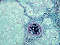
In a first, researchers at the University of Iowa and the University of Missouri (MU) have developed a pig model for cystic fibrosis (CF) that appears to closely mimic the disease in human infants. The striking similarities between disease manifestations in the CF piglets and human newborns with CF suggest that this new model will help improve understanding of the disease and may also speed discovery of new treatments. The study is published in the Sept. 26 issue of Science.
CF is a common hereditary disease that affects multiple organ systems, including the intestines, pancreas, and lung. Mice with CF-causing mutations have helped researchers learn more about this disease, however, differences in physiology and biology mean that mice with CF mutations do not develop many of the typical symptoms that affect humans with CF.
"Lack of a better model has hampered our ability to answer long-standing questions in CF," explained Christopher Rogers, Ph.D., a former postdoctoral fellow in internal medicine at the UI Roy J. and Lucille A. Carver College of Medicine, and one of the study's lead authors. "The CF pig provides a unique opportunity to study one of the most common genetic diseases, and we hope to translate this new knowledge into better therapies and preventions."
In addition to Rogers, co-lead authors of the study were David Stoltz, M.D., Ph.D., UI assistant professor of internal medicine, and David Meyerholz, D.V.M., Ph.D., UI assistant professor of pathology.
The senior study author was Michael Welsh, M.D., UI professor of internal medicine and molecular physiology and biophysics, who holds the Roy J. Carver Chair of Internal Medicine and Physiology and Biophysics. Welsh also is a Howard Hughes Medical Institute investigator.
CF occurs when a person inherits two mutated copies of the CFTR gene leading to loss of ion channel function that adversely affects many organs. To create the CF pigs, the researchers used gene targeting to disrupt one copy of the normal gene in pig cells. They then cloned these altered cells to produce pigs with only one good copy of the gene. Like human CF-carriers, these animals did not show disease symptoms. The pigs were then bred naturally, and about one in four of the piglets were born with two disrupted copies of the gene.
The researchers established that piglets lacking CFTR have the abnormal ion channel activity that is a hallmark of CF disease. They also showed that the CF piglets develop the same disease characteristics that are commonly seen in newborn humans with CF, including a bowel obstruction known as meconium ileus, which often is the first sign of CF in humans. The pigs also have an abnormal pancreas, liver, and gall bladder, similar to CF patients.
"Thus far, the clinical, physiological and age-related appearance of disease in the pigs, as well as the organs involved, mimic CF seen in people," Stoltz said.
A primary cause of death and disability in patients with CF is lung disease. However, many questions remain about how infection and inflammation leads to lung damage. In the study, the lungs of the newborn CF pigs appeared similar to the lungs of their normal littermates and had no sign of infection or inflammation, possibly shedding some initial insight on the process. As the CF pigs mature and are exposed to airborne bacteria and viruses, the researchers hope to learn more about how and why lung disease develops in patients with CF.
"Researchers can now begin to study the disease progression as it is happening, something not possible in humans," Meyerholz said.
The research team included UI scientists from internal medicine, pediatrics, surgery and periodontics. In addition, Randall Prather, Ph.D., the distinguished professor of reproductive biology at MU College of Agriculture, Food and Natural Resources, and his colleagues were part of the research team.
The study was funded in part by grants from the National Heart, Lung, and Blood Institute and National Institute of Diabetes and Digestive and Kidney Disease of the National Institutes of Health, Food for the 21st Century and the Cystic Fibrosis Foundation.
Gene Variant Boosts Risk of Fatty Liver Disease
Researchers at UT Southwestern Medical Center have found that individuals who carry a specific form of the gene PNPLA3 have more fat in their livers and a greater risk of developing liver inflammation.
They also found that Hispanics are more likely to carry the gene variant responsible for higher liver-fat content than African-Americans and Caucasians.
The new findings, published online today in the journal Nature Genetics, provide a gene-based explanation for the results of a 2004 UT Southwestern-led study that determined that the propensity to develop nonalcoholic fatty liver disease differs among ethnic groups, with a higher percentage of Hispanics developing the disorder than African-Americans or Caucasians.
“A single variation in the PNPLA3 gene was strongly associated with hepatic fat content, even after adjusting for other factors, such as obesity, diabetes status and alcohol intake,” said senior study author Dr. Helen Hobbs, director of the Eugene McDermott Center for Human Growth and Development and an investigator for the Howard Hughes Medical Institute at UT Southwestern.
“Sequence variations in this gene explain much of the increased propensity of Hispanics to accumulate excess liver fat,” she said.
Nonalcoholic fatty liver disease (NAFLD) is the most common form of liver disease in Western countries, and its incidence is growing. Previous UT Southwestern research has shown that it may affect as many as one-third of adults in America.
“The gene variations we have identified might provide a way to predict who is most at risk for developing fatty liver disease and liver injury in response to environmental stresses such as obesity or infection,” said Dr. Jonathan Cohen, professor of internal medicine in the McDermott Center and one of the authors of the study.
NAFLD results from the accumulation of triglycerides in the liver and is associated with metabolic disorders such as insulin resistance, obesity, diabetes and high cholesterol – many of the conditions that contribute to heart disease. It can also lead to liver inflammation, cirrhosis and liver cancer. Approximately 10 percent of liver transplants performed in the U.S. are for cirrhosis related to NAFLD, according to the researchers.
Treatments for NAFLD include weight loss, exercise, reducing alcohol intake and improved diabetes control.
“Knowing who is at increased risk of developing liver disease could aid physicians in encouraging their patients to make lifestyle changes or take other preventive measures to help mitigate their underlying genetic risk for the disorder,” said Dr. Cohen, holder of the C. Vincent Prothro Distinguished Chair in Human Nutrition Research.
The new findings come out of the Dallas Heart Study, an 8-year-old groundbreaking investigation of cardiovascular disease that involves a large multiethnic group of participants. The $12 million study investigates the links between genetics, lifestyle and the risks for heart disease.
As part of the Dallas Heart Study, more than 3,500 individuals from Dallas County provided blood samples in 2000 for DNA isolation and other tests. Each participant also underwent multiple body scans with magnetic resonance imaging and computed tomography to examine the heart and other organs. Along with discovering new genetic ties to differences in blood levels of cholesterol and triglycerides, the researchers have used this information to identify new drug targets for the prevention and treatment of heart disease.
Data-gathering for the new study on fatty liver disease took advantage of a unique aspect of the Dallas Heart Study. Researchers with the study were the first to analyze hepatic fat in a large population using a technique called proton magnetic resonance spectroscopy – the most sensitive and quantitative noninvasive imaging technique available to measure the amount of fat in the liver. With this technique, they screened more than 2,100 individuals across multiple ethnicities. They then correlated that data with DNA tests from the same people and found the link to the PNPLA3 gene.
The next step in the research is to investigate how the various forms of the PNPLA3 gene affect lipid metabolism.
Other researchers from the McDermott Center involved with the study were lead author Dr. Stefano Romeo, a postdoctoral research fellow; Dr. Alexander Pertsemlidis, assistant professor; Dr. Chao Xing, assistant professor of clinical sciences; and Julia Kozlitina, a graduate student participating in a joint program between Southern Methodist University and UT Southwestern. Researchers from Perlegen Sciences, Lawrence Berkeley National Laboratory and the UT Health Science Center at Houston also were involved.
The research was funded by the Donald W. Reynolds Foundation, the National Institutes of Health and the Department of Energy.
Monday, May 12, 2008
Modeling psychiatric disorders in mice
One of the molecular causes of the behavioral and cognitive deficits observed in mice with a small chromosomal deletion has been identified, according to a study published online this week in Nature Genetics. The corresponding deletion in the human genome gives rise to a range of psychiatric disorders, and accounts for approximately 1–2% of cases of schizophrenia in the general population.
Deletion of a small region on chromosome 22 is associated with anxiety, depression, attention-deficit hyperactivity disorder, autism, and deficits in working memory. Approximately 30% of the individuals carrying such a deletion eventually develop schizophrenia.
Maria Karayiorgou, Joseph Gogos and colleagues generated a model in which the corresponding region was deleted in the mouse genome. Mice carrying a single deletion show a range of behavioral and cognitive deficits that mimic some aspects of the human syndrome. Of particular interest is the increased expression of precursors of the small regulatory RNAs known as microRNAs in the brains of the mutant mice. The authors went on to show that loss of one of genes in the deleted region, Dgcr8, is responsible for this increased abundance of precursors, as the normal role of Dgcr8 is to process them into mature microRNAs. By generating mice that have only one copy of Dgcr8, the authors showed that this mutation by itself results in at least some of the deficits observed in mice with the deletion of the surrounding region.
Although the specific downstream targets of altered microRNA expression in the brain are not yet known, the authors suggest these findings could have general implications for understanding the genetic basis of psychiatric disorders.
Author contacts:
Maria Karayiorgou (Columbia University Medical Center, New York, NY, USA)
Tel: +1 212 568 4189; E-mail: mk2758@columbia.edu
Joseph Gogos (Columbia University Medical Center, New York, NY, USA)
Tel: +1 212 305 0744; E-mail: jag90@columbia.edu
Stem-cell regeneration of the breast relies on adhesion
Interaction of basal stem cells in the breast with their cellular environment is crucial for their function, and helps towards the regeneration of the mammary glands during pregnancy, reports a paper online this week in Nature Cell Biology.
The basal stem cells of the breast are enriched in proteins called integrins that mediate contact with the extracellular matrix surrounding the cells. Marina Glukhova and colleagues show that expression of beta 1 integrin in the basal cells is essential for the regenerative potential of these stem cells and for proper development of the mammary gland during pregnancy.
Deletion of the beta 1 integrin gene from the basal layer of mouse mammary tissue led to an abnormal ductal branching pattern in the mammary gland during pregnancy, which results in the regenerative potential of the mammary tissue stem cells being abolished, leading to a dysfunctional gland. In basal stem cells lacking beta 1 integrin, cells divide abnormally and this results in the altered branching pattern.
The environment that surrounds most stem cells, the stem cell niche, is known to be important for a number of stem cells. However, our understanding of stem cell niches remains patchy. These findings establish a central role of direct interaction between basal stem cells and their extracellular matrix in the maintenance of the mammary stem-cell population.
Author contact:
Marina Glukhova (CNRS-Institut Curie Research, Paris, France)
Tel: +33 1 42 34 63 33; E-mail: Marina.Glukhova@curie.fr
The origins of the modern tomato
Scientists have identified a genetic mutation that acts as a major contributor to the extreme fruit size associated with the modern tomato, according to a study.
Modern cultivated tomatoes produce fruit as much as 1,000 times larger than their wild progenitors. One clear reason for the increase in tomato size during its domestication is the increased number of carpels – organs – which determines the final number of compartments in the fruit.
Steven Tanksley and colleagues crossed lines of tomatoes with either high or low compartment number, and carried out genetic mapping studies to identify the gene or genes responsible for the variation in carpel number. They identified an insertion of 6–8 kilobases in a gene they call fas only in the tomatoes with high compartment number. Expression of the gene is reduced in the developing flower buds in tomatoes carrying the insertion. A survey of 30 different lines of cerasiforme, the wild form of tomato thought to be related to the smaller progenitors, showed that none carried the insertion.
As the insertion is found exclusively in modern cultivated tomatoes, the authors suggest that the mutation occurred recently in tomato domestication, and then spread rapidly as a result of selection for larger fruit.
Author contact:
Steven Tanksley (Cornell University, Ithaca, NY, USA)
Tel: +1 607 255 1673; E-mail: sdt4@cornell.edu
Friday, May 09, 2008
The planets: Mysteries surrounding the ‘butterscotch’ planet’s equator
Saturn, the second largest planet in the Solar System, is easily spotted because of the brightness of the rings around its equator. Two companion papers in this week’s Nature report on features of its atmosphere, one using data collected by the Cassini mission and the other from over two decades of ground-based observations.
The equatorial stratospheres of Earth and Jupiter oscillate more or less periodically on timescales of about two and four years, respectively. By analysing infrared observations from the Cassini probe, Thierry Fouchet and colleagues discovered that Saturn has an equatorial oscillation like Earth's and Jupiter's, as well as a mid-latitude subsidence that may be associated with the equatorial motion. Glenn Orton and co-workers’ ground-based observations of Saturn's stratospheric emission reveal a similar oscillation.
The period of the oscillation is approximately 15 terrestrial years, which is roughly half of Saturn's year, suggesting the influence of seasonal forcing —rather like the Earth's semi-annual oscillation.
CONTACT
Glenn Orton (Jet Propulsion Laboratory, Pasadena, CA, USA) Author paper [3]
Tel: +1 818 354 2460; E-mail: Glenn.Orton@jpl.nasa.gov
Thierry Fouchet (Observatoire de Paris, Meudon, France) Author paper [4]
Tel: +33 1 45 07 71 11; E-mail: Thierry.Fouchet@obspm.fr
Timothy Dowling (University of Louisville, KY, USA) N&V author
Tel: +1 502 852 3927; E-mail: dowling@louisville.ed
Genomes: Is it a bird, is it a mammal…?
The duck-billed platypus (Ornithorhynchus anatinus) is a truly unique animal, and its fascinating genome. Platypuses are monotremes with almost no close relatives alive on earth. Scientists just had to take a look at that genome, and now an international collaboration of researchers report its sequencing and analysis.
Famously considered a hoax when sent from Australia to European researchers in the nineteenth century, the platypus is an amalgam of reptilian, mammalian and unique characteristics that provide clues to the function and evolution of all mammalian genomes. Sequencing of the platypus genome has helped to uncover the following: the origins of genomic imprinting in vertebrates; platypus venom proteins were co-opted independently from the same gene families that provided reptile venom; milk protein genes are conserved; and immune gene family expansions are directly related to platypus biology. As well as providing an invaluable resource for comparative genomics, the sequence will be important for monotreme conservation.
CONTACT
Wesley Warren (Washington University School of Medicine, St Louis, MO, USA)
Tel: +1 314 286 1899; E-mail: wwarren@wustl.edu
Jennifer Marshall Graves (Australian National University, Canberra, Australia)
Tel: +61 261 252 492; E-mail: jenny.graves@anu.edu.au
Ewan Birney (The European Bioinformatics Institute, Cambridge, UK)
E-mail: birney@ebi.ac.uk
Monday, May 05, 2008
Developmental genetics: Starting out on the road to maleness
Growing up male is a genetic lifestyle decision — early in embryonic development, genes on the Y chromosome activate the development of specialized cells that ultimately become the testes. Without this ‘on switch’ for maleness, the developing gonads become ovaries by default and the embryo develops as a female.A study of mice now shows how the developing gonads start out on this road to maleness. In a paper geneticists Robin Lovell-Badge and Ryohei Sekido describe how a gene called Sry, carried on the Y chromosome, boosts another gene elsewhere in the genome that in turn governs the development of sperm-producing cells called Sertoli cells — a crucial component of the testes.By studying gene expression patterns in developing mouse embryos, the researchers deduced that Sry produces a protein that combines with another protein, steroidogenic factor 1 (SF1). This complex then binds to a DNA region that boosts the expression of another gene, Sox9, which controls a host of genes involved in sperm development.Elucidating this pathway not only reveals how maleness develops from the ‘default’ female developmental pathway; defects in this process may also explain how people who are genetically ‘male’ or ‘female’ end up developing the ‘wrong’ sexual anatomy.
contact:
Robin Lovell-Badge (National Institute for Medical Research, London, UK)Tel: +44 20 8816 2126;
Genetic susceptibility to obesity
Scientists have discovered genetic variants that increase the risk of obesity and insulin resistance in the general population, according to two studies published online this week in Nature Genetics. Until now only one locus (FTO) has been associated convincingly with increased risk of obesity.A consortium of investigators led by Mark McCarthy, Ines Barroso and Nicholas Wareham analyzed the genomes of more than 90,000 individuals and found that a variant near MC4R, encoding the melanocortin-4 receptor, increases susceptibility to obesity. Previous studies had shown that the melanocortin-4 receptor is expressed in neurons in the hypothalamus and is a key regulator of food intake and energy expenditure. Although it is unclear how this variant affects MC4R expression or function, the fact that mutations in the gene are known to cause rare cases of severe childhood obesity lends confidence to the association.In a separate study, Jaspal Kooner and colleagues carried out a genome-wide scan of several thousand individuals of Indian Asian or European ancestry, and identified a variant near MC4R as increasing risk of obesity and insulin resistance. The risk variant was more frequent in individuals of Indian Asian ancestry, which the authors suggest may account for the increased burden of obesity in Indian Asians.Author contacts:Mark McCarthy (University of Oxford, UK) Author paper [7]Tel: +44 1865 857 298; E-mail: mark.mccarthy@drl.ox.ac.ukInês Barroso (Wellcome Trust Sanger Institute, Hinxton, UK) Co-author paper [7]Tel: +44 1223 495 341; E-mail: ib1@sanger.ac.ukNicholas Wareham (Addenbrooke’s Hospital, Cambridge, UK) Co-author paper [7]Tel: +44 1223 330 315; E-mail: nick.wareham@mrc-epid.cam.ac.ukJaspal Kooner (Imperial College London, UK) Author paper [8]Tel: +44 20 8383 4751; E-mail: j.kooner@imperial.ac.uk
Obesity: Your number of fat cells stays constant in adulthood
number of fat cells, or adipocytes, in your body remains more or less constant throughout adulthood, a new study finds। The discovery suggests that the difference in the number of fat cells between lean and obese people is established in childhood, and then persists for life।The number of fat cells remains constant even in formerly obese adults who have lost significant amounts of weight, report researchers led by Kirsty Spalding, who studied fat samples from liposuction and abdominal reconstruction surgery in lean and obese volunteers। This reflects the fact that the level of obesity is determined by a combination of the number and size of fat cells, which can grow or shrink as fat from food is deposited in them।Although fat cell numbers remain constant during adulthood, this is a dynamic process of death and replenishment, Spalding and her colleagues report. Fat cells are replaced at the same rate that they die — roughly 10% every year. The researchers made their discovery by studying levels of radioactive isotopes in fat cells from people who had lived through the brief period of Cold War nuclear bomb testing from 1955 to 1963. People whose fat cells were deposited before the onset of testing nevertheless incorporated radioactive matter after, showing that their fat cells were being replenished.The fact that fat cells are constantly dying and being replaced could potentially offer an opportunity to develop new anti-obesity therapies, the researchers suggest.
Author contact:
Kirsty Spalding (Karolinska Institute, Stockholm, Sweden)Tel: +46 70 437 1542; E-mail:
kirsty.spalding@cmb.ki.se
Thursday, May 01, 2008
The Hunt for the Kill Switch
The U.S. Department of Defense wants to know if chip makers are building remotely accessible kill switches into high-end microprocessors. These days, the U.S. military consumes only about 1 percent of the world's integrated circuits, and offshoring has begun to raise some alarms about the safety of the chips in the military's most mission-critical electronics.
Recognizing an enormous vulnerability, the DOD recently launched an extremely ambitious program to verify the integrity of the electronics that will underpin future additions to its arsenal. In December, the DOD's advanced-research wing released details about a three-year initiative it calls the Trust in Integrated Circuits program. The findings from the program could give the military--and defense contractors who make sensitive microelectronics like the weapons systems for the F-35--a guaranteed method of determining whether their chips have been compromised with a kill switch.
But how exactly would you kill an integrated switch, and for what purpose? In "The Hunt for the Kill Switch," IEEE Spectrum's Sally Adee reports on the methods that could kill a chip, the possible consequences, and the methods being devised to verify the Pentagon's most important microchips.(NewsWise)
Researchers Grow Heart and Blood Cells from Reprogrammed Skin Cells
Stem cell researchers at UCLA were able to grow functioning cardiac cells using mouse skin cells that had been reprogrammed into cells with the same unlimited properties as embryonic stem cells.
The finding is the first to show that induced pluripotent stem cells or iPS cells, which don’t involve the use of embryos or eggs, can be differentiated into the three types of cardiovascular cells needed to repair the heart and blood vessels.
The discovery could one day lead to clinical trials of new treatments for people who suffer heart attacks, have atherosclerosis or are in heart failure, said Dr. Robb MacLellan, a researcher at the Eli and Edythe Broad Center of Regenerative Medicine and Stem Cell Research and senior author of the study. Researchers also were able to differentiate the iPS cells into several types of blood cells, which may one day aid in treating blood diseases and in bone marrow transplantation.
“I believe iPS cells address many of the shortcomings of human embryonic stem cells and are the future of regenerative medicine,” said MacLellan, an associate professor of cardiology and physiology. “I’m hoping that these scientific findings are the first step towards one day developing new therapies that I can offer my patients. There are still many limitations with using iPS cells in clinical studies that we must overcome, but there are scientists in labs across the country working to address these issues right now.”
The study, which brought together stem cell and cardiology researchers at UCLA, appears online May 1, 2008 in the journal Stem Cells. The article can be accessed at www.stemcells.com/papbyrecent.dtl.
Last June, UCLA stem cell researchers were among several scientific teams that were the first to reprogram mouse skin cells into cells resembling embryonic stem cells, which have the ability to become every cell type found in the body. MacLellan and his team used UCLA’s iPS cells in their study.
Although iPS cells are believed to be very similar to embryonic stem cells, further study needs to be done to confirm their differentiation potential. MacLellan’s study proved that iPS cells can be induced into becoming cardiovascular cells, an important step in the confirmation process.
“Theoretically, iPS cells are able to differentiate into 220 different cells types,” said Dr. Miodrag Stojkovic, co-editor of Stem Cells. “For the first time, scientists from UCLA were able to induce the differentiation of mouse iPS cells into functional heart cells.”
In MacLellan’s study, the iPS cells were cultured on a protein matrix known to direct embryonic stem cells into differentiating into cardiovascular progenitor cells, immature heart cells that can give rise to mature cardiac cells that perform different functions. The progenitor cells were then isolated from the other iPS cells that did not differentiate using a protein marker called KDR, a growth factor receptor expressed on the surface of the progenitor cells.
Once isolated, the cardiovascular progenitor cells were coaxed into becoming cardiomyoctyes, or mature heart muscle cells that control heartbeat, endothelial cells, which form rudimentary blood vessels, and vascular smooth muscle cells, the specialized cells that line blood vessel walls. Once mature, the cardiomyocytes beat in the Petri dish.
Studies are under way now at UCLA to determine if human iPS cells behave the same way as the mouse cells behave. If they do, the time may come when a person could use their own skin cells to create individualized iPS cell lines to provide cells for cardiac repair and regeneration, MacLellan said.
It is vital to be able to grow and isolate progenitor, or partially differentiated, cells that can create the three types of cardiac cells for potential clinical use. When embryonic stem cells are injected directly into the heart in animal models, they create tumors because the cells differentiate not only into cardiac cells but into other cells found in the human body as well. Likewise, using embryonic stem cells garnered from other sources than the patient could result in rejection of the injected cells.
The use of iPS cells may solve those problems. If the iPS cells come from the patient, rejection should not be an issue. Additionally, the use of cells that are already partially transformed into specific cardiac cell types may prevent tumor growth. The use of iPS cells also sidesteps the controversy some associate with deriving pluripotent stem cells from embryos or eggs, MacLellan said.
“Our hope is that, based on this work in mice, we can show that similar cardiovascular progenitor cells can be found in human iPS cells and, using a similar strategy, that we can isolate the progenitor cells and differentiate them into the cells types found in the human heart,” MacLellan said.
The stem cell center was launched in 2005 with a UCLA commitment of $20 million over five years. A $20 million gift from the Eli and Edythe Broad Foundation in 2007 resulted in the renaming of the center. With more than 150 members, the Eli and Edythe Broad Center for Regenerative Medicine and Stem Cell Research is committed to a multi-disciplinary, integrated collaboration of scientific, academic and medical disciplines for the purpose of understanding adult and human embryonic stem cells. The institute supports innovation, excellence and the highest ethical standards focused on stem cell research with the intent of facilitating basic scientific inquiry directed towards future clinical applications to treat disease. The center is a collaboration of the David Geffen School of Medicine, UCLA’s Jonsson Cancer Center, the Henry Samueli School of Engineering and Applied Science and the UCLA College of Letters and Science
Saturday, April 12, 2008
Blood Vessels: the Pied Piper for Growing Nerve Cells
Researchers at Johns Hopkins have discovered that blood vessels in the head can guide growing facial nerve cells with blood pressure controlling proteins. The findings, which suggest that blood vessels throughout the body might have the same power of persuasion over many nerves, are published this week in Nature.
“We’re excited to have stumbled across another family of proteins that can tell a growing nerve which way to grow,” says David Ginty, Ph.D., a professor of neuroscience at Hopkins and investigator of the Howard Hughes Medical Institute. “But the really interesting thing is that the nerves appear to use blood vessels as guideposts to direct their growth in one of several possible directions.”
The research team studied in mice a group of about 15,000 nerve cells known as the superior cervical ganglia, or SCG, which extend projections that innervate various structures in the head including the eyes, mouth and salivary glands. The SCG sits in a Y-like branching point of the blood vessel in the neck that supplies the head with blood, the carotid artery. In the developing embryo, nerve projections grow out of the SCG and grow along one of the two branches of the carotid artery; the nerves that grow along the internal carotid innervate the eyes and mouth among other head structures, and those that grow along the external carotid innervate the salivary glands.
To figure out how nerve cells “choose” to grow along the external carotid artery to innervate the salivary glands, the team looked for genes that appear to be preferentially turned on in the external carotid, and off in the internal carotid. Says Ginty, “There’s only two directions they can go and we wanted to know if they choose their direction or if the decision to go one way or the other is random.”
They found one gene that is expressed preferentially in the external carotid, a gene that makes the blood pressure regulating protein, endothelin, active. “It comes as no surprise that something critical for regulating the cardiovascular system in the adult also is used for directing nerve growth in the developing embryo,” says Ginty. “The genome is limited and nature has figured out a way to use things over and over again for unrelated functions.”
Further examination of the arteries in mouse embryos confirmed that endothelin is found only in the external carotid. To confirm that the nerve cell projections grow toward endothelin, the researchers removed SCGs and grew each one next to an endothelin-soaked bead. Checking on them three days later, the team found that nerves from the SCGs had grown towards the beads. To be certain that endothelin directs nerve growth in the living animal, the researchers then looked in mice that had the endothelin gene removed. Sure enough, these mice had no nerves growing along their external carotid arteries.
The team then wondered if all growing nerves in the SCG can respond to endothelin. So they looked for the endothelin receptors in SCG nerves and found only a subset of SCG nerves make endothelin receptors and concluded that those nerves somehow already had been chosen to respond to the endothelin made by the external carotid.
“How do these nerve cells know which target organ they’re supposed to innervate when they all come from the same progenitor?” asks Ginty. “This is what we’re going to study next.”
The research was funded by the National Institutes of Health and the Howard Hughes Medical Institute.
Authors on the paper are Takako Makita and Ginty of Hopkins; Henry Sucov of the University of Southern California; Cheryl Gariepy of the University of Michigan; and Masashi Yanagisawa of the University of Texas Southwestern Medical Center.
“Black Gold Agriculture” May Revolutionize Farming, Curb Global Warming
Fifteen hundred years ago, tribes people from the central Amazon basin mixed their soil with charcoal derived from animal bone and tree bark. Today, at the site of this charcoal deposit, scientists have found some of the richest, most fertile soil in the world. Now this ancient, remarkably simple farming technique seems far ahead of the curve, holding promise as a carbon-negative strategy to rein in world hunger as well as greenhouse gases.
At the 235th national meeting of the American Chemical Society, scientists report that charcoal derived from heated biomass has an unprecedented ability to improve the fertility of soil — one that surpasses compost, animal manure, and other well-known soil conditioners.
They also suggest that this so-called “biochar” profoundly enhances the natural carbon seizing ability of soil. Dubbed “black gold agriculture,” scientists say this “revolutionary” farming technique can provide a cheap, straight-forward strategy to reduce greenhouse gases by trapping them in charcoal-laced soil.
“Charcoal fertilization can permanently increase soil organic matter content and improve soil quality, persisting in soil for hundreds to thousands of years,” Mingxin Guo, Ph.D., and colleagues report. In what they describe as a “new and pioneering” ACS report — the first systematic investigation of soil improvement by charcoal fertilization — Guo found that soils receiving charcoal produced from organic wastes were much looser, absorbed significantly more water and nutrients and produced higher crop biomass. The authors, with Delaware State University, say “the results demonstrate that charcoal amendment is a revolutionary approach for long-term soil quality improvement.”
Soil deterioration from depletion of organic matter is an increasingly serious global problem that contributes to hunger and malnutrition. Often a result of unsustainable farming, overuse of chemical fertilizers and drought, the main weapons to combat the problem —compost, animal manure and crop debris — decompose rapidly.
“Earth’s soil is the largest terrestrial pool of carbon,” Guo said. “In other words, most of the earth’s carbon is fixed in soil.” But if this soil is intensively cultivated by tillage and chemical fertilization, organic matter in soil will be quickly decomposed into carbon dioxide by soil microbes and released into the atmosphere, leaving the soil compacted and nutrient-poor.
Applying raw organic materials to soil only provides a temporary solution, since the applied organic matter decomposes quickly. Converting this unutilized raw material into biochar, a non-toxic and stable fertilizer, could keep carbon in the soil and out of the atmosphere, says Guo.
“Speaking in terms of fertility and productivity, the soil quality will be improved. It is a long-term effect. After you apply it once, it will be there for hundreds of years,” according to Guo. With its porous structure and high nutrient- and water-holding capabilities, biochar could become an extremely attractive option for commercial farmers and home gardeners looking for long-term soil improvement.
The researchers planted winter wheat in pots of soil in a greenhouse. Some pots were amended with two percent biochar, generated from readily available ingredients like tree leaves, corn stalk and wood chips. The other pots contained ordinary soil.
The biochar-infused soil showed vastly improved germination and growing rates compared to regular soil. Guo says that even a one-percent charcoal treatment would lead to improved crop yield.
Guo is “positive” that this ground-breaking farming technique can help feed countries with poor soil quality. “We hope this technology will be extended worldwide,” says Guo.
“The production of current arable land could be significantly improved to provide more food and fiber for the growing populations. We want to call it the second agricultural revolution, or black gold revolution!”
He suggests that charcoal production has been practiced for at least 3000 years. But until now, nobody realized that this charcoal could improve soil fertility until archaeologists stumbled on the aforementioned Amazonian soil several years ago.
Biochar production is straightforward, involving a heating process known as pyrolysis. First, organic residue such as tree leaves and wood chips is packed into a metal container and sealed. Then, through a small hole on top, the container is heated and the material burns. The raw organic matter is transformed into black charcoal. Smokes generated during pyrolysis can also be collected and cooled down to form bio-oil, a renewable energy source, says Guo.
In lieu of patenting biochar, Guo says he is most interested in extending the technology into practice as soon as possible. To that end, his colleagues at Delaware State University are investigating a standardized production procedure for biochar. They also foresee long-term field studies are needed to validate and demonstrate the technology. Guo noted that downsides of biochar include transportation costs resulting from its bulk mass and a need to develop new tools to spread the granular fertilizer over large tracts of farmland.
The researchers are about to embark on a five-year study on the effect of “black gold” on spinach, green peppers, tomatoes and other crops. They seek the long-term effects of biochar fertilization on soil carbon changes, crop productivity and its effect of the soil microorganism community.
“Through this long-term work, we will show to people that biochar fertilization will significantly change our current conventional farming concepts,” says Guo.
The American Chemical Society — the world’s largest scientific society — is a nonprofit organization chartered by the U.S. Congress and a global leader in providing access to chemistry-related research through its multiple databases, peer-reviewed journals and scientific conferences. Its main offices are in Washington, D.C., and Columbus, Ohio.
Subscribe to:
Posts (Atom)
