Wednesday, June 24, 2009


First Direct Visualization of Memory Formation in the Brain

FINDINGS: UCLA and McGill University researchers have, for the first time, “photographed” a memory in the making. The study clarifies one of the ways in which connections in the brain between nerve cells, called synapses, can be changed with experience. The phenomenon is called “synaptic plasticity,” and is the foundation for how we learn and remember. As we learn, the memories are stored in changes in the strength and/or number of synaptic connections between nerve cells in our brain. Long lasting changes in synaptic connections are required for long-term memories, and the persistence of these changes requires new gene expression. This is the first study to use fluorescent imaging to directly visualize protein synthesis at individual synapses during learning related synaptic plasticity.

IMPACT: Understanding how synapses can change with experience is critical to understanding behavioral plasticity, and to understanding diseases in which learning and experience-dependent behaviors are impaired. Such diseases include mental retardation, Alzheimer’s disease, as well as anxiety and mood disorders. It also can elucidate potential strategies for improving normal cognition and behavioral plasticity.

JOURNAL: The research appears in the June 19 edition of the journal Science.

AUTHORS: Senior author Kelsey Martin, associate professor of psychiatry and biological chemistry; Dan Ohtan Wang, Sang Mok Kim, Yali Zhao, Hongik Hwang, Satoru K. Miura, all of UCLA; and Wayne S. Sossin, McGill University.

HOW: The researchers used sensory and motor neurons from the sea slug Aplysia Californica that can form connections in culture. The neurons were stimulated with serotonin, which strengthens the synapses, and allowed them to detect new protein synthesis—the making of a memory— using a “translational reporter,” a fluorescent protein that can be easily detected and tracked.

MORE: This is the first study to directly visualize protein synthesis at individual synapses during a long-lasting form of synaptic plasticity. The studies revealed an exquisite level of control over the specificity of regulation of new protein synthesis. “While this was not really surprising to us given the complexity of information processing in the brain,” said Martin, “visualizing the process of protein synthesis at individual synapses, and beginning to discern the elegance of its regulation, leaves us, as biologists, with a wonderful sense of awe.”

Funding: This study was funded by the National Institutes of Health, the WM Keck Foundation, and the Canadian Institutes of Health Research. The authors report no conflict of interest.

Friday, June 19, 2009

Bacteria Can Plan Ahead

Bacteria can anticipate a future event and prepare for it, according to new research at the Weizmann Institute of Science. In a paper that appeared in the June 17, 2009 issue of Nature, Prof. Yitzhak Pilpel, doctoral student Amir Mitchell, and research associate Dr. Orna Dahan of the Institute’s Molecular Genetics Department, together with Prof. Martin Kupiec and Gal Romano of Tel Aviv University, examined microorganisms living in environments that change in predictable ways. Their findings show that these microorganisms’ genetic networks are hard-wired to “foresee” what comes next in the sequence of events and begin responding to the new state of affairs before its onset.

E. coli bacteria, for instance, which normally cruise harmlessly down the digestive tract, encounter a number of different environments on their way. In particular, they find that one type of sugar – lactose – is invariably followed by a second sugar – maltose – soon afterward. Pilpel and his team in the Molecular Genetics Department checked the bacteria’s genetic response to lactose and found that, in addition to the genes that enable it to digest lactose, the gene network for utilizing maltose was partially activated. When they switched the order of the sugars, giving the bacteria maltose first, there was no corresponding activation of lactose genes, implying that bacteria have naturally “learned” to get ready for a serving of maltose after a lactose appetizer.

Another microorganism that experiences consistent changes is wine yeast. As fermentation progresses, sugar and acidity levels change, alcohol levels rise, and the yeast’s environment heats up. Although the system was somewhat more complicated than that of E. coli, the scientists found that when the wine yeast feel the heat, they begin activating genes for dealing with the stresses of the next stage. Further analysis showed that this anticipation and early response is an evolutionary adaptation that increases the organism’s chances of survival.

Ivan Pavlov first demonstrated this type of adaptive anticipation, known as a conditioned response, in dogs in the 1890s. He trained the dogs to salivate in response to a stimulus by repeatedly ringing a bell before giving them food. In the microorganisms, says Pilpel, “evolution over many generations replaces conditioned learning, but the end result is similar.” “In both evolution and learning,” says Mitchell, “the organism adapts its responses to environmental cues, improving its ability to survive.” Romano: “This is not a generalized stress response, but one that is precisely geared to an anticipated event.” To see whether the microorganisms were truly exhibiting a conditioned response, Pilpel and Mitchell devised a further test for the E. coli based on another of Pavlov’s experiments. When Pavlov stopped giving the dogs food after ringing the bell, the conditioned response faded until they eventually ceased salivating at its sound. The scientists did something similar, using bacteria grown by Dr. Erez Dekel, in the lab of Prof. Uri Alon of the Weizmann Institute’s Molecular Cell Biology Department, in an environment containing the first sugar, lactose, but not following it up with maltose. After several months, the bacteria had evolved to stop activating their maltose genes at the taste of lactose, only turning them on when maltose was actually available.

“This showed us that there is a cost to advanced preparation, but that the benefits to the organism outweigh the costs in the right circumstances,” says Pilpel. What are those circumstances? Based on the experimental evidence, the research team created a sort of cost/benefit model to predict the types of situations in which an organism could increase its chances of survival by evolving to anticipate future events. The researchers are already planning a number of new tests for their model, as well as different avenues of experimentation based on the insights they have gained.

Pilpel and his team believe that genetic conditioned response may be a widespread means of evolutionary adaptation that enhances survival in many organisms – one that may also take place in the cells of higher organisms, including humans. These findings could have practical implications, as well. Genetically engineered microorganisms for fermenting plant materials to produce biofuels, for example, might work more efficiently if they gained the genetic ability to prepare themselves for the next step in the process.

Prof. Yitzhak Pilpel’s research is supported by the Ben May Charitable Trust and Madame Huguette Nazez, Paris, France.

The Weizmann Institute of Science in Rehovot, Israel, is one of the world's top-ranking multidisciplinary research institutions. Noted for its wide-ranging exploration of the natural and exact sciences, the Institute is home to 2,600 scientists, students, technicians, and supporting staff. Institute research efforts include the search for new ways of fighting disease and hunger, examining leading questions in mathematics and computer science, probing the physics of matter and the universe, creating novel materials, and developing new strategies for protecting the environment.
Key Found to How Tumor Cells Invade the Brain in Childhood Cancer

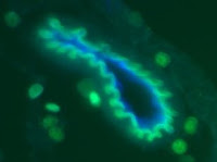
Despite great strides in treating childhood leukemia, a form of the disease called T-cell acute lymphoblastic leukemia (T-ALL) poses special challenges because of the high risk of leukemic cells invading the brain and spinal cord of children who relapse. Now, a new study in the June 18, 2009, issue of the journal Nature by scientists at NYU School of Medicine reveals the molecular agents behind this devastating infiltration of the central nervous system. The finding may lead to new drugs that block these agents and thus lower the risk of relapse.

T-ALL, a blood-borne cancer in which the bone marrow makes too many lymphocytes, or white blood cells, strikes several hundred children and adolescents in the U.S. annually. While greater than 90% percent go into remission through a combination of chemotherapy and radiation, up to one third of this group end up relapsing. These patients are at particular risk for tumor cells to invade the brain and spinal cord, and to prevent this all patients receive chemotherapy injections into the central nervous system and in some cases cranial irradiation—approaches that cause dangerous side effects, including secondary tumors and potentially permanent cognitive and developmental deficits.

“In general, T-cell acute lymphoblastic leukemia is treatable with chemotherapy and radiation,” said Ioannis Aifantis, PhD, associate professor of pathology and co-director of the Cancer Stem Cell Program at the NYU Cancer Institute, who led the new study. “But you have a very high rate of relapse. And after the relapse, it is not treatable because the cancer occurs in tricky places like the central nervous system,” said Dr. Aifantis, who is also an Early Career Scientist at the Howard Hughes Medical Institute.

“We are very proud of this research and very excited about the potential implications for new therapeutic approaches to prevent or reduce the spread of leukemic cells into the central nervous system,” said Vivian S. Lee MD, PhD, MBA, the vice dean for science, senior vice president and chief scientific officer of NYU Langone Medical Center.

In the new study, Dr. Aifantis and his colleagues found that a key protein receptor embedded on the outer surface of leukemic cells is responsible for infiltrating the brain and spinal cord. “What we have found is that leukemic cells over-express this receptor.” said Dr. Aifantis, “If you knock out this receptor, these cells will not go to the brain under any circumstances.”

Previous research had strongly implicated a famous gene regulator called Notch1 in the progression of T-ALL. The Notch1 gene (a mutated version gives fruit flies notched wings) is an oncogene, or cancer-causing gene, in humans. Certain kinds of mutations in this gene have been found in nearly half of all T-ALL patients, and current estimates suggest that the gene’s regulatory influence might be implicated in nearly 90 percent of all T-ALL cases.

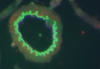
For their new study, Dr. Aifantis and his colleagues first introduced overactive forms of Notch1 into mice. As a result, the mice developed leukemia and the leukemic cells efficiently infiltrated the inner layers of the membrane covering the brain. “What happens is that the leukemic cells get into the cerebrospinal fluidthat protects our brain and spine, where they fill up the space and they can affect brain function, either by secreting chemicals and toxic factors or even by simple pressure,” Dr. Aifantis said.

His team then examined an array of other mouse genes to identify candidates that might fall under the regulatory spell of Notch1 to promote the brain and spinal cord infiltration. The screen revealed a promising gene for a protein named CCR7, which is embedded on the surface of lymphocytes. This chemokine receptor, as it’s known, normally senses and responds to small chemical attractants called chemokines, which act like recruitment signals for lymphocytes to converge on a specific site during the body’s response to infection or injury. In leukemia, however, these lymphocytes proliferate abnormally.


CCR7 was already known as a key player in normal lymphocyte migration and as a binding partner of two chemokines named CCL19 and CCL21. Previous studies had implicated these protein interactions in the metastasis of other tumors such as melanomas and breast cancers. Dr. Aifantis’s team also discovered that the gene for CCR7 was overactive in four of five T-ALL cell lines derived from human patients, bolstering suspicions that it played a central role in the disease. Conversely, a mutation that knocked out Notch1 also led to dramatically reduced CCR7 levels.

To characterize CCR7’s potential role in T-ALL, the researchers used two sets of mice: one in which the receptor was turned on, and a second in which it was turned off. When the team delivered an identical number of human-derived leukemic cells to both sets of mice, those with the CCR7 chemokine receptor turned off lived almost twice as long. Using bioluminescent imaging, the researchers quickly understood why: animals with the active CCR7 receptor had many more tumors. Tellingly, the T-ALL cells had infiltrated the brain and spinal cord of those mice.

Further experiments suggested that when healthy mice received leukemic cells in which the gene for CCR7 had been turned off, the cells could not migrate to the brain even though they reached other body tissues. As a result, the mice survived significantly longer than counterparts with an active copy of the gene. On the other hand, introducing a normal version of the same gene to mice otherwise lacking it was enough to recruit leukemic cells to the brain and spine.

“We wanted to determine whether CCR7 by itself was sufficient for entry into the central nervous system and that’s what this experiment shows,” Dr. Aifantis said. “By changing one specific gene, you now have your function back.”

Finally, the researchers identified the small protein that acted as the “come hither” signal for the CCR7 protein receptors. One candidate, CCL21, was undetectable in leukemic mice. But a second, CCL19, appeared in tiny veins of the brain near the infiltrating tumor cells. When the researchers introduced leukemic cells carrying a gene for CCR7 to mice that naturally lacked the CCL19 chemokine, the mice survived longer, suggesting that their increased life spans might be due to a disrupted interaction of the two proteins. The leukemic cells had no trouble infiltrating other tissue like the lymph nodes, but were completely incapable of infiltrating the brains of CCL19-deficient mice, the researchers report.

“Perhaps there are antibodies or small molecules that can block the interaction between these two proteins or reduce their interactions,” Dr. Aifantis said, “and hopefully that could be used as a type of prophylactic treatment to prevent a relapse in the central nervous system among patients who have already been treated for leukemia.” Such a treatment, he said, could prove a good alternative to the intensive and often poorly tolerated radiation and chemotherapy now used to try to block such a relapse.

The study was led by Dr. Silvia Buonamici, a post-doctoral fellow in the laboratory of Dr. Aifantis in the Department of Pathology and the NYU Cancer Institute, and in the Helen L. and Martin S. Kimmel Stem Cell Center at NYU Langone Medical Center. Other study investigators are; Thomas Trimarchi, Maria Grazia Ruocco, Linsey Reavie, Severine Cathelin, Yevgeniy Lukyanov, Jen-Chieh Tseng, Filiz Sen, Mengling Li, Elizabeth Newcomb, Jiri Zavadil, Daniel Meruelo, Sherif Ibrahim, David Zagzag, and Michael L. Dustin from NYU Langone Medical Center; Brenton G. Mar, Apostolos Klinakis, and Argiris Efstratiadis from Columbia University Medical Center; Eric Gehrie and Jonathan S. Bromberg from Mount Sinai School of Medicine; and Martin Lipp from the Max Delbrück Center for Molecular Medicine in Berlin.

The study was supported by grants from the National Institutes of Health, the American Cancer Society, the Dana Foundation, The Chemotherapy Foundation, the Alex’s Lemonade Stand Foundation, the Lauri Strauss Leukemia foundation, the G&P Foundation, an NYU School of Medicine Molecular Oncology and Immunology training grant, the American Society of Hematology, the Juvenile Diabetes Research Foundation, the National Cancer Institute, a gift from the Berrie Foundation, and a fellowship from the Jane Coffin Childs Memorial Fund for Medical Research.

About NYU Langone Medical Center
Located in New York City, NYU Langone Medical Center is one of the nation's premier centers of excellence in health care, biomedical research, and medical education. For over 168 years, NYU physicians and researchers have made countless contributions to the practice and science of health care. Today the Medical Center consists of NYU School of Medicine, including the Smilow Research Center, the Skirball Institute of Biomolecular Medicine, and the Sackler Institute of Graduate Biomedical Sciences; the three hospitals of NYU Hospitals Center, Tisch Hospital, a 705-bed acute-care general hospital, Rusk Institute of Rehabilitation Medicine, the first and largest facility of its kind, and NYU Hospital for Joint Diseases, a leader in musculoskeletal care; and such major programs as the NYU Cancer Institute, the NYU Child Study Center, and the Hassenfeld Children's Center for Cancer and Blood Disorders.

About NYU Cancer Institute
The NYU Cancer Institute is an NCI-designated cancer center. Its mission is to discover the origins of human cancer and to use that knowledge to eradicate the personal and societal burden of cancer in our community, the nation and the world. The center and its multidisciplinary team of experts provide access to the latest treatment options and clinical trials along with a variety of programs in cancer prevention, screening, diagnostics, genetic counseling and supportive services. For additional information, please visit: www.nyuci.org.

Tuesday, June 09, 2009

Confusion About Sugars

Three top researchers corrected inaccuracies and misunderstandings concerning high fructose corn syrup's impact on the American diet. They also examined how the United States Department of Agriculture (USDA) considers this sweetener in light of the upcoming 2010 Dietary Guidelines for Americans in a session, High Fructose Corn Syrup: Sorting Myth from Reality, at the Institute of Food Technologists (IFT) Annual Meeting in Anaheim, California.


"Contrary to its name, high fructose corn syrup is essentially a corn sugar," stated sweetener expert John S. White, Ph.D., president of White Technical Research. "Recent marketing claims that sugar is healthier than high fructose corn syrup are misleading to consumers."


"By every parameter yet measured in human beings, high fructose corn syrup and sugar are identical. This is not surprising since high fructose corn syrup and sugar are metabolized the same by the body, have the same level of sweetness and the same number of calories per gram," noted James M. Rippe, M.D., cardiologist and biomedical sciences professor at the University of Central Florida.


"This is a marketing issue, not a metabolic issue," stated David Klurfeld, Ph.D., national program leader for human nutrition in USDA's Agricultural Research Service and editor of the June 2009 Journal of Nutrition supplement, "The State of the Science on Dietary Sweeteners Containing Fructose," in response to recent reformulations by manufacturers of products that once contained high fructose corn syrup. "The real issue is not high fructose corn syrup. It's that we've forgotten what a real serving size is. We have to eat less of everything," he noted.


Increased Caloric Intake, Not a Single Sweetener, the Likely Cause of Obesity


Fructose-containing sweeteners -- such as sugar, invert sugar, honey, fruit juice concentrates, and high fructose corn syrup -- are essentially interchangeable in composition, calories, and metabolism. Replacing high fructose corn syrup in foods with other fructose-containing sweeteners will provide neither improved nutrition nor a meaningful solution to the obesity crisis, according to Dr. White. "In light of similarities in composition, sweetness, energy content, processing, and metabolism, claims that such sweetener substitutions bring nutritional benefit to children and their families appear disingenuous and misguided," White says.


Growing Body of Evidence


The American Medical Association helped put to rest a common misunderstanding about high fructose corn syrup and obesity, stating that "high fructose syrup does not appear to contribute to obesity more than other caloric sweeteners." Even former critics of high fructose corn syrup dispelled myths and distanced themselves from earlier speculation about the sweetener's link to obesity in a comprehensive scientific review published in the December 2008 American Journal of Clinical Nutrition.